. 2008 Oct 1;232(1):25-32.
doi: 10.1016/j.taap.2008.05.024. Epub 2008 Jun 4.

LY294002 inhibits glucocorticoid-induced COX-2 gene expression in cardiomyocytes through a phosphatidylinositol 3 kinase-independent mechanism

Affiliations

LY294002 inhibits glucocorticoid-induced COX-2 gene expression in cardiomyocytes through a phosphatidylinositol 3 kinase-independent mechanism

Haipeng Sun et al. Toxicol Appl Pharmacol. .

Abstract

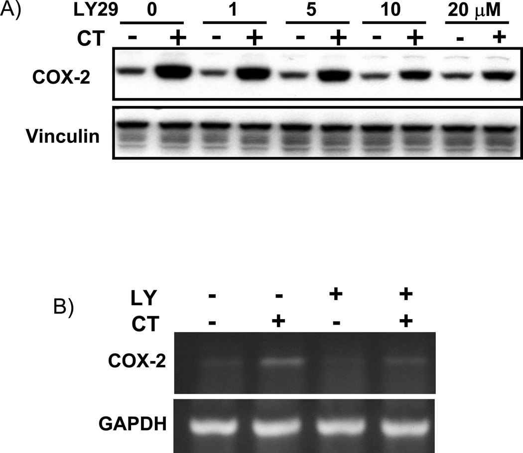
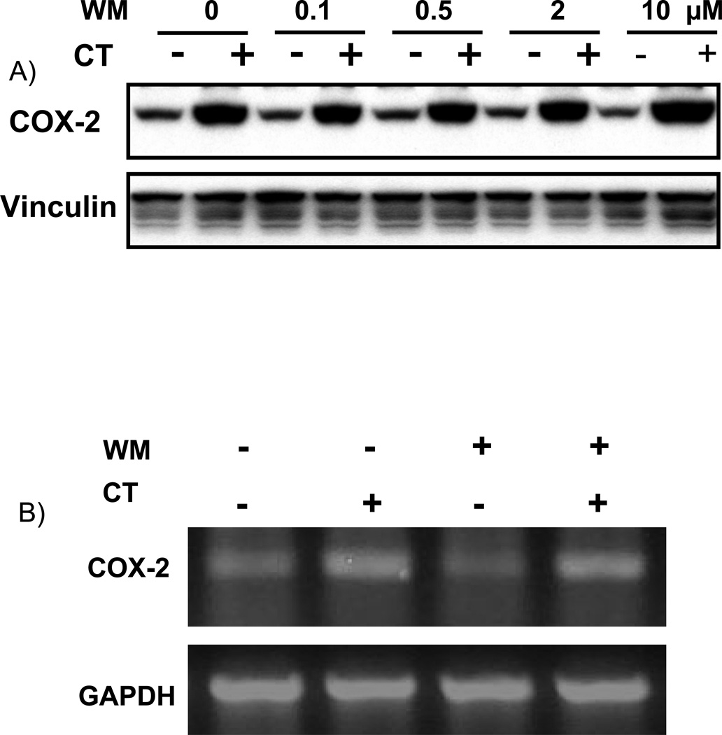
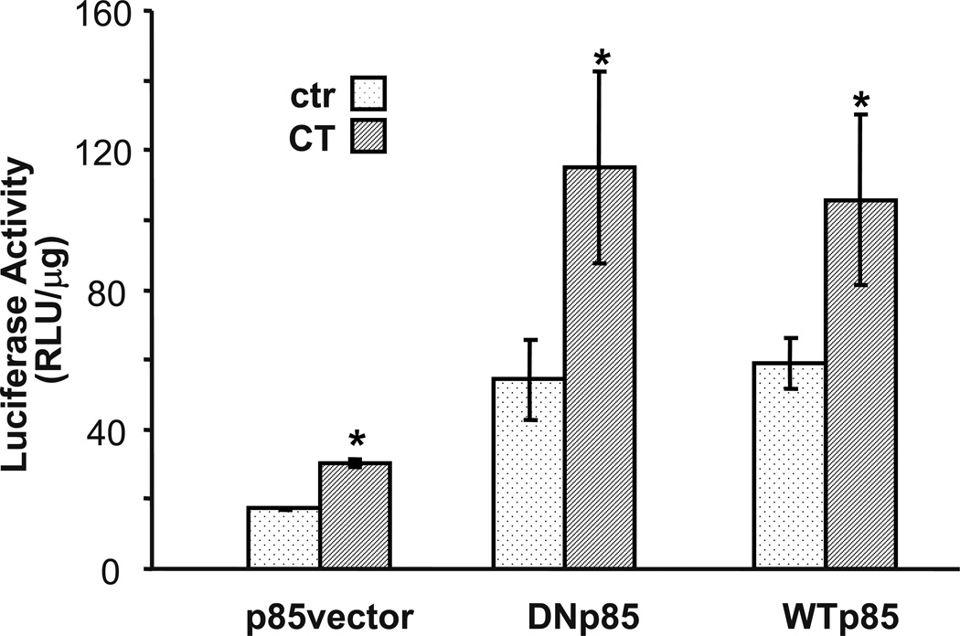
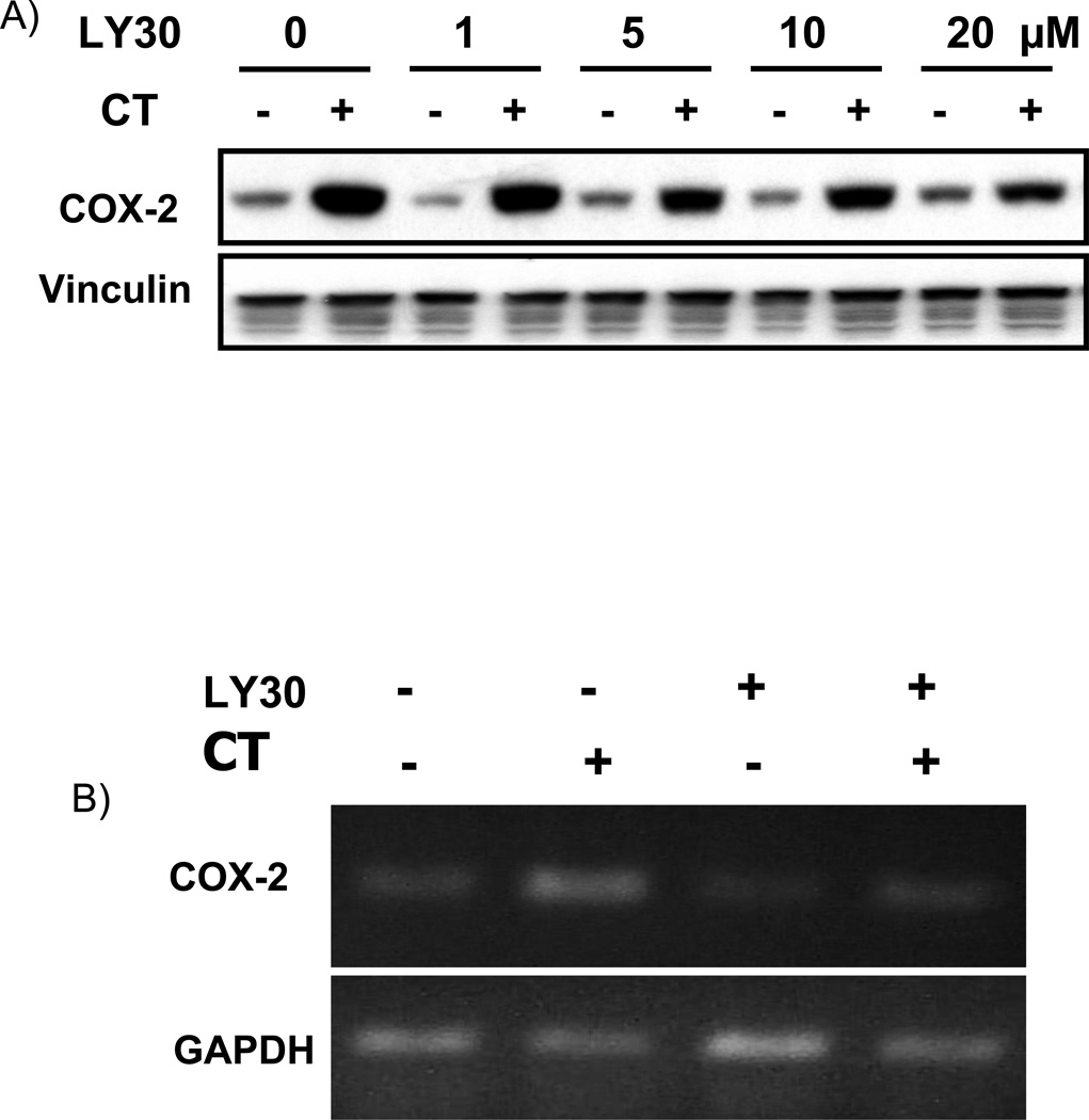
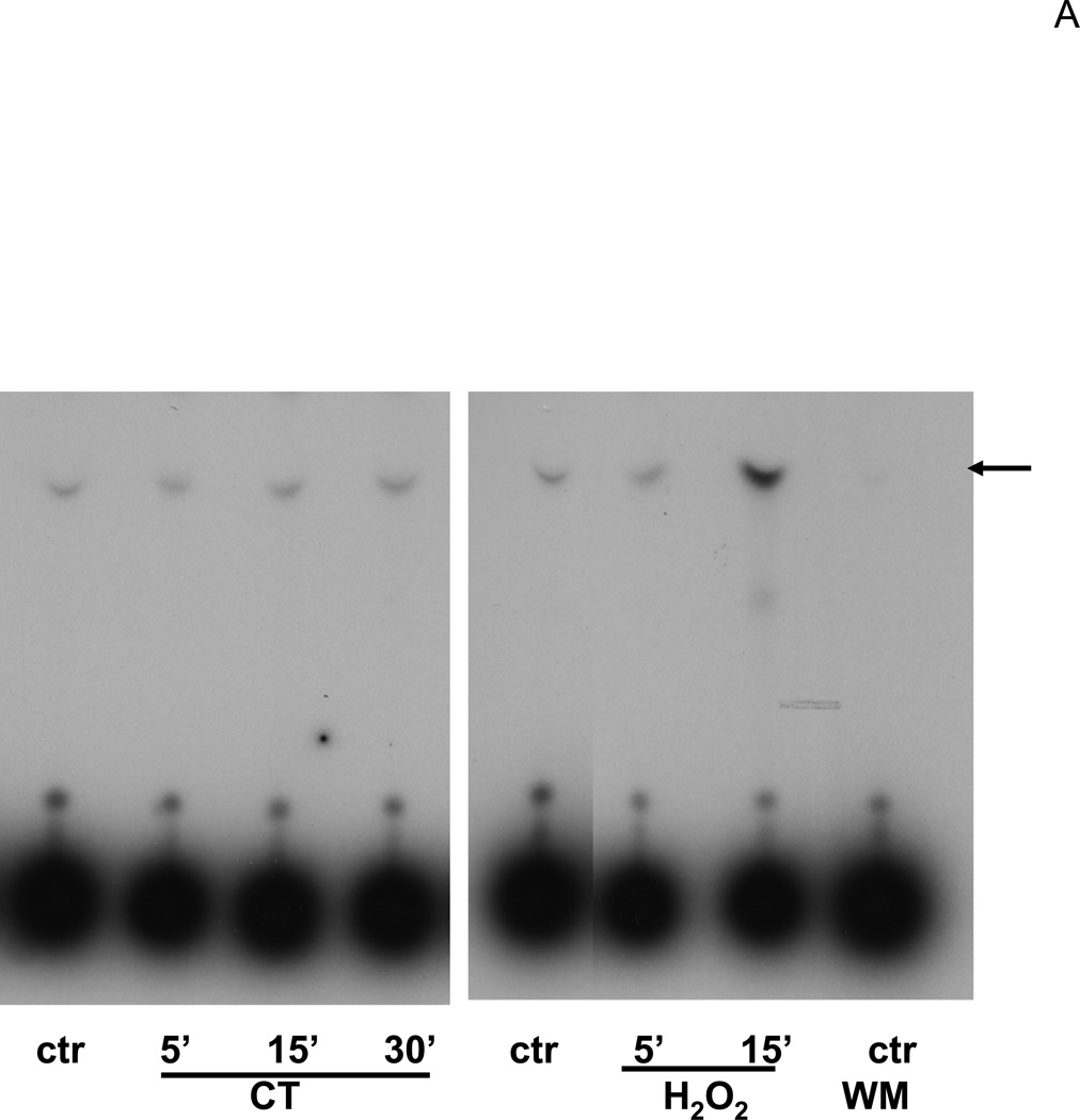
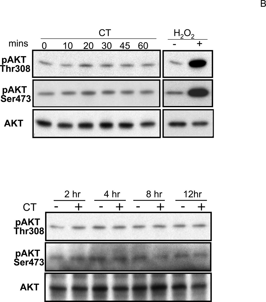
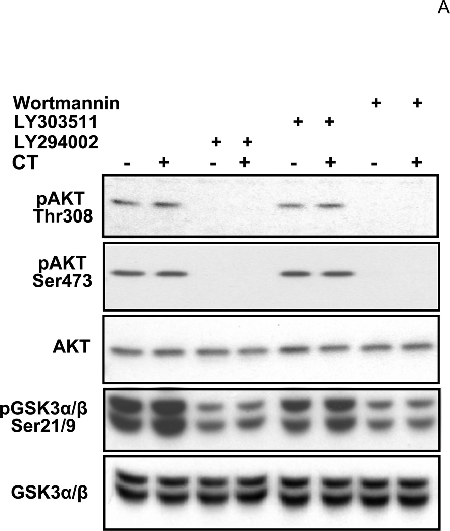
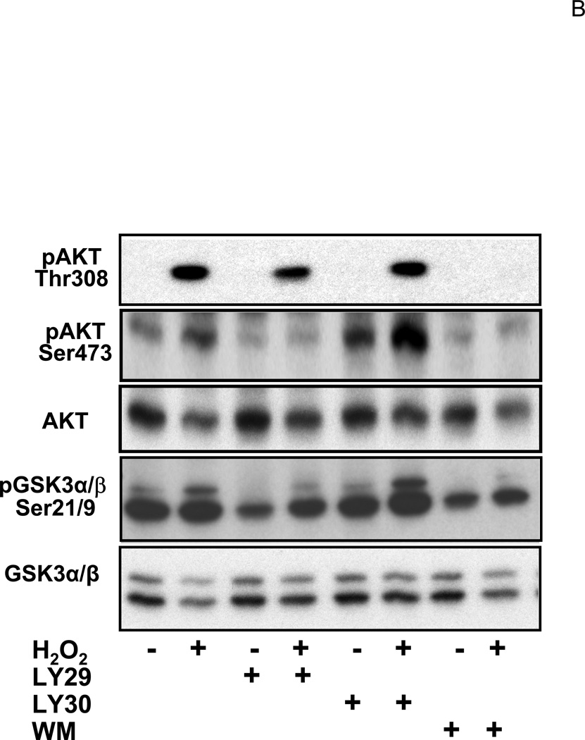
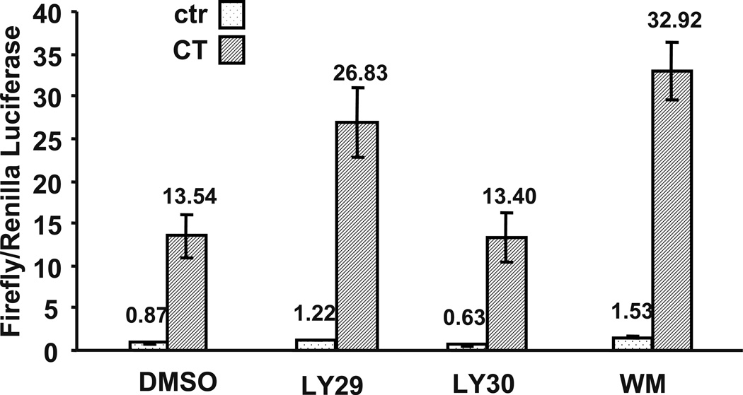
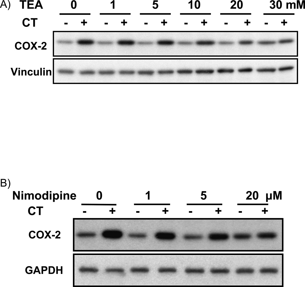
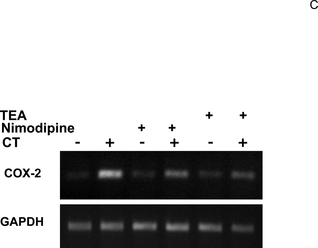
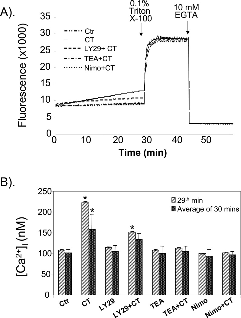
Glucocorticoids induce COX-2 expression in rat cardiomyocytes. While investigating whether phosphatidylinositol 3 kinase (PI3K) plays a role in corticosterone (CT)-induced COX-2, we found that LY294002 (LY29) but not wortmannin (WM) attenuates CT from inducing COX-2 gene expression. Expression of a dominant-negative mutant of p85 subunit of PI3K failed to inhibit CT from inducing COX-2 expression. CT did not activate PI3K/AKT signaling pathway whereas LY29 and WM decreased the activity of PI3K. LY303511 (LY30), a structural analogue and a negative control for PI3K inhibitory activity of LY29, also suppressed COX-2 induction. These data suggest PI3K-independent mechanisms in regulating CT-induced COX-2 expression. LY29 and LY30 do not inhibit glucocorticoid receptor transactivity. Both compounds have been reported to inhibit Casein Kinase 2 activity and modulate potassium and calcium levels independent of PI3K, while LY29 has been reported to inhibit mammalian Target of Rapamycin (mTOR), and DNA-dependent Protein Kinase (DNA-PK). Inhibitor of Casein Kinase 2 (CK2), mTOR or DNA-PK failed to prevent CT from inducing COX-2 expression. Tetraethylammonium (TEA), a potassium channel blocker, and nimodipine, a calcium channel blocker, both attenuated CT from inducing COX-2 gene expression. CT was found to increase intracellular Ca(2+) concentration, which can be inhibited by LY29, TEA or nimodipine. These data suggest a possible role of calcium instead of PI3K in CT-induced COX-2 expression in cardiomyocytes.


Figures

Figure 1
Figure 1. LY294002 dose dependent inhibition of CT-induced COX-2 elevation
Cardiomyocytes were pretreated with LY29 at the dose indicated (A) or 10 µM (B) 60 mins before treatment of 1 µM CT for 4 hours (A) or 2 hours (B). COX-2 was measured by Western blot (A) or RT-PCR (B) with vinculin or GAPDH as a loading control. Alternatively, cardiomyocytes were transfected with rat COX-2 promoter luciferase vector (C). Transfected cells with or without 60-mins pretreatment of 10 µM LY29 were treated with 1 µM CT for 12 hours before luciferase assay (C). The activity of luciferase is expressed as the Relative Light Unit (RLU) per µg protein (C). The data represent one of three independent experiments. An asterisk (*) indicates significant difference (p<0.05) as judged by the Student's t test when the means of CT treated groups were compared to that of the control in three experiments (C).
Figure 2
Figure 2. Wortmannin failed to inhibit CT-induced COX-2 elevation
Cardiomyocytes were pretreated with WM at the dose indicated (A) or 2 µM (B) 60 mins before treatment of 1 µM CT for 4 (A) or 2 hours (B). COX-2 was measured by Western blot (A) or RT-PCR (B) with vinculin or GAPDH as a loading control. Transfected cardiomyocytes with or without 60-mins pretreatment of 2 µM WM were treated with 1 µM CT for 12 hours before harvesting for measurements of luciferase activity, which is expressed as the RLU per µg protein (C). The data represent one of three independent experiments. An asterisk (*) indicates significant difference (p<0.05) as judged by the Student's t test when the means of CT treated groups were compared to that of the control from three independent experiments (C).
Figure 3
Figure 3. Dominant negative mutant of PI3K failed to prevent COX-2 induction
Cardiomyocytes were transfected with rat COX-2 promoter reporter vector along with empty vector, wild type or dominant-negative p85 expression vector. Transfected cells were incubated with or without 1 µM CT for 12 hours before luciferase activity assay. The activity of luciferase is expressed as the RLU per µg protein. The data show one experiment representative of three. An asterisk (*) indicates significant difference (p<0.05) as judged by the Student's t test when the means of CT treated groups were compared to that of the control from three independent experiments.
Figure 4
Figure 4. LY303511 attenuates CT-induced COX-2 elevation
Cardiomyocytes were pretreated with LY30 at the dose indicated (A) or 10 µM (B) 60 mins before treatment of 1 µM CT for 4 hours (A) or 2 hours (B). COX-2 was measured by Western blot (A) or RT-PCR (B) with vinculin (A) or GAPDH as a loading control (B).
Figure 5
Figure 5. CT failed to activate PI3K/AKT signaling pathway
Cardiomyocytes were treated with 1 μM CT for indicated time before harvesting for measurements of PI3K activity (A), Thr308 or Ser473 phospho AKT, or total AKT (B). H2O2 treatment (100 μM, 15 mins for A or 30 mins for B) was included as a positive control. In the last lane, wortmannin (WM) was added to the reaction mixture to inhibit PI3K in vitro and indicate the specific product of PI3K activity assay (A). The data represent one of three independent experiments.
Figure 6
Figure 6. Effects of LY294002, LY303511 and Wortmannin on PI3K/AKT pathway
Cardiomyocytes were treated with 1µM CT for 4hr before harvesting for measurements of Thr308 or Ser473 phospho AKT, total AKT, Ser21/9 phospho GSK3 α/β or total GSK3 α/β. LY29 (20 µM), LY30 (20 µM) or WM (2 µM) was added to cells 60 mins prior to CT. The data represent one of three independent experiments.
Figure 7
Figure 7. Effects of LY294002, LY303511, and Wortmannin on glucocorticoid receptor activation
Cardiomyocytes were transfected with MMTV-luc and TK-Renilla luciferase vector. Cells were treated with LY29 (10 µM), LY30 (10 µM) or WM (2 µM) for 60 mins before 24 hr treatment of 1 µM CT. Cells were harvested for duel luciferase assay and MMTV firefly luciferase activity was normalized to TK-Renilla luciferase activity. The bars represent the ratio of MMTV firefly luciferase versus TK-Renilla luciferase. The fold of induction by CT treatment is indicated above the bars. An asterisk (*) indicates significant difference (p<0.05) as judged by the Student's t test when the means of CT treated groups were compared to that of the control from three independent experiments.
Figure 8
Figure 8. Effect of TEA or nimodipine on CT induced COX-2 expression
Cardiomyocytes were pretreated 60 mins with TEA (A) or nimodipine (nimo, B) at the dose indicated, or with TEA (30 mM) or nimodipine (20 µM) (C), before treatment of 1 µM CT for 4 hours. COX-2 was measured by Western blot (A,B) or RT-PCR (C) with with vinculin (A, B) or GAPDH (C) as a loading control. The data represent one of three independent experiments.
Figure 9
Figure 9. Efffect of LY29, TEA and nimodipine on CT induced [Ca2+]i increase
Cardiomyocytes seeded in a 12-well plate were placed in HEPES buffer for fluo-4 AM dye loading and fluorescence measurement using a microplate reader (BioTek ELX800) as described in the Method. The instrument measures fluorescence intensity every 7 seconds. The fluorescence reflecting [Ca2+]i changes for control, CT or CT plus inhibitors are shown (A). The [Ca2+]i for each sample was calculated as average ± standard deviation based on 258 measurements during 30 mins interval or 9 measurements during the last minute (29th min) before Triton X-100 addition (B). The data are from one experiment representative of three independent experiments. An asterisk (*) indicates significant difference (p<0.05) as judged by the Student's t test when the means of CT treated groups were compared to that of the control.

Similar articles

Cited by

References

    1. Adcock IM, Ito K, Barnes PJ. Glucocorticoids: Effects on Gene Transcription. Proc. Am. Thorac. Soc. 2004;1:247–254. - PubMed
    1. Adderley SR, Fitzgerald DJ. Oxidative Damage of Cardiomyocytes Is Limited by Extracellular Regulated Kinases 1/2-mediated Induction of Cyclooxygenase-2. J. Biol. Chem. 1999;274:5038–5046. - PubMed
    1. Alessi DR, Andjelkovic M, Caudwell B, Cron P, Morrice N, Cohen P, Hemmings BA. Mechanism of activation of protein kinase B by insulin and IGF-1. EMBO J. 1996;15:6541–6551. - PMC - PubMed
    1. Bachelor MA, Cooper SJ, Sikorski ET, Bowden GT. Inhibition of p38 Mitogen-Activated Protein Kinase and Phosphatidylinositol 3-Kinase Decreases UVB-Induced Activator Protein-1 and Cyclooxygenase-2 in a SKH-1 Hairless Mouse Model. Mol. Cancer Res. 2005;3:90–99. - PubMed
    1. Bolli R, Shinmura K, Tang XL, Kodani E, Xuan YT, Guo Y, Dawn B. Discovery of a new function of cyclooxygenase (COX)-2: COX-2 is a cardioprotective protein that alleviates ischemia/reperfusion injury and mediates the late phase of preconditioning. Cardiovasc. Res. 2002;55:506–519. - PMC - PubMed

Publication types

MeSH terms