. 2008 Oct 3;382(2):434-47.
doi: 10.1016/j.jmb.2008.07.027. Epub 2008 Jul 16.

Structure of the myristylated human immunodeficiency virus type 2 matrix protein and the role of phosphatidylinositol-(4,5)-bisphosphate in membrane targeting

Affiliations

Structure of the myristylated human immunodeficiency virus type 2 matrix protein and the role of phosphatidylinositol-(4,5)-bisphosphate in membrane targeting

Jamil S Saad et al. J Mol Biol. .

Abstract

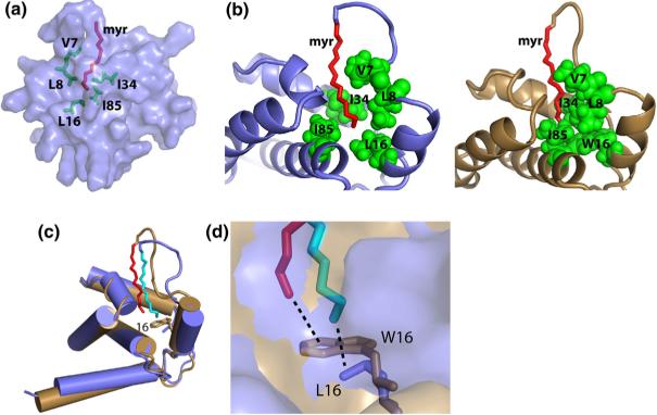
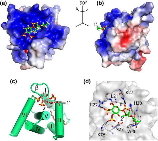
During the late phase of retroviral replication, newly synthesized Gag proteins are targeted to the plasma membrane (PM), where they assemble and bud to form immature virus particles. Membrane targeting by human immunodeficiency virus type 1 (HIV-1) Gag is mediated by the PM marker molecule phosphatidylinositol-(4,5)-bisphosphate [PI(4,5)P(2)], which is capable of binding to the matrix (MA) domain of Gag in an extended lipid conformation and of triggering myristate exposure. Here, we show that, as observed previously for HIV-1 MA, the myristyl group of HIV-2 MA is partially sequestered within a narrow hydrophobic tunnel formed by side chains of helices 1, 2, 3, and 5. However, the myristate of HIV-2 MA is more tightly sequestered than that of the HIV-1 protein and does not exhibit concentration-dependent exposure. Soluble PI(4,5)P(2) analogs containing truncated acyl chains bind HIV-2 MA and induce minor long-range structural changes but do not trigger myristate exposure. Despite these differences, the site of HIV-2 assembly in vivo can be manipulated by enzymes that regulate PI(4,5)P(2) localization. Our findings indicate that HIV-1 and HIV-2 are both targeted to the PM for assembly via a PI(4,5)P(2)-dependent mechanism, despite differences in the sensitivity of the MA myristyl switch, and suggest a potential mechanism that may contribute to the poor replication kinetics of HIV-2.


Figures

Fig. 1
Fig. 1
(a) Overlay of 2D 1H–15N HSQC spectra collected for HIV-2 myr(−)MA (black) and myr(+)MA (red) proteins (500 μM, 35 °C). Significant chemical shift changes occur for residues that are affected by the positioning of the myr group. (b) Representative sedimentation profiles obtained for myr(−)MA (black) and myr(+)MA (red) proteins (26,000 rpm, 20 °C, 110 μM). For both proteins, sedimentation profiles fit best to monomeric species.
Fig. 2
Fig. 2
Three-dimensional 13C-edited/12C-double-half-filtered NOE data obtained for HIV-2 myr(+)MA showing unambiguously assigned intermolecular NOEs between the myristate group and key residues of a 13C-labeled protein sample (myristate group is not 13C labeled). Continuous and dashed lines denote 1H–12C breakthrough doublets and NOE peaks, respectively.
Fig. 3
Fig. 3
Stereoviews showing the best-fit backbone superpositions of the 20 refined structures calculated for HIV-2 myr(−)MA (top) and myr(+)MA (bottom) proteins. The myristate group is shown in red. The last 15 residues of the protein are not shown because they are disordered.
Fig. 4
Fig. 4
A representative structure of HIV-2 myr(+)MA (slate) and comparison with the HIV-1 myr(+)MA protein (sand). (a) Semitransparent surface representation of MA showing the penetration of the myr group (red sticks) and interactions with the side chains of Val7, Leu8, Leu16, Ile34, and ILe85 (green sticks). (b) Cartoon representation of the HIV-2 and HIV-1 myr(+)MA proteins comparing the sequestration of the myristate group (red) in the hydrophobic cavity formed by residues Val7, Leu8, Leu16, Ile34, and Ile85 (green spheres). (c) Superimposition of representative structures of HIV-2 and HIV-1 myr(+)MA. Myristate groups of HIV-2 and HIV-1 myr(+)MA proteins are packing against Leu16 and Trp16, respectively. NMR data revealed that helix 6 is flexible for both proteins. (d) An expanded view of the protein core of HIV-2 and HIV-1 myr(+)MA showing the myristate packing against side chains of Leu16 and Trp16, respectively.
Fig. 5
Fig. 5
Comparison of the HIV-2 myr(−)MA structure with the myristate-exposed HIV-1 myr(+)MA structure. The packing of helix 1 is stabilized by a buried His(+)— Glu(−) salt bridge in the HIV-1 protein, whereas the packing is stabilized by a buried His−Lys(+) salt bridge in the HIV-2 protein.
Fig. 6
Fig. 6
(a) Overlay of 2D 1H–15N HSQC spectra upon titration of HIV-2 myr(+)MA with di-C4-PI(4,5)P2 [60 μM, 35 °C; di-C4-PI(4,5)P2/MA = 0:1 (black), 1:1 (red), 2:1 (green), 4:1 (blue), 8:1 (magenta), and 16:1 (cyan)]. (b) 1H and 15N NMR chemical shift titration data, which fit to 1:1 binding isotherms (Kd = 143±16 μM). (c) Representative 13C-edited/12C-double-half-filtered NOE data showing unambiguously assigned intermolecular NOEs between residues Leu21, Ser77, and di-C4-PI(4,5)P2. Dashed lines denote intermolecular NOE peaks.
Fig. 7
Fig. 7
Structure of the HIV-2 myr(+)MA/di-C4-PI(4,5)P2 complex. (a,b) Interactions between di-C4-PI(4,5)P2 (sticks) and MA (colored according to electrostatic surface potential) showing the 2′-fatty acid inserting in a preexisting cleft and the inositol ring packing against a basic patch of the protein. (c) PI(4,5)P2 binding to the β-II–V cleft. (d) A network of interactions implicated in PI(4,5)P2 binding.
Fig. 8
Fig. 8
Disruption of PI(4,5)P2 inhibits HIV-1 and HIV-2 particle production. HeLa cells were transfected with HIV-1 (pNL4−3) or HIV-2 (pROD10) molecular clones alone (−) or were co-transfected at a 1:2 DNA ratio with vectors expressing 5ptaseIV, the Δ1 deletion mutant of 5ptaseIV, or Arf6/Q67L. Transfected cells were metabolically radiolabeled with [35S]Met/Cys for 2 h. Cell and viral lysates were prepared and immunoprecipitated with HIV-Ig (for HIV-1 samples) or with the anti-p27gag monoclonal antibody R1C7 (for HIV-2 samples). Gag proteins were quantified by phosphorimager analysis. Virus release efficiency was calculated as the amount of virion-associated Gag as a fraction of total (cell plus virion) Gag.
Fig. 9
Fig. 9
Disruption of PI(4,5)P2 induces a relocalization of HIV-1 and HIV-2 assembly to intracellular vesicles. HeLa cells were transfected with HIV-1 (pNL4−3) or HIV-2 (pROD10) molecular clones alone (−) or were co-transfected with vectors expressing 5ptaseIVor Arf6/Q67L. Transfected cells were fixed and examined by thin-section transmission EM. Scale bars represent 100 nm unless otherwise indicated.

References

    1. Lemey P, Pybus OG, Wang B, Saksena NK, Salemi M, Vandamme A-M. Tracing the origin and history of the HIV-2 epidemic. Proc. Natl Acad. Sci. USA. 2003;100:6588–6592. - PMC - PubMed
    1. Poulsen AG, Aaby P, Larsen O, Jensen H, Naucler A, Lisse IM, et al. 9-year HIV-2-associated mortality in an urban community in Bissau, West Africa. Lancet. 1997;349:911–914. - PubMed
    1. Schim van der Loeff MF, Jaffar S, Aveika AA, Sabally S, Corrah T, Harding E, et al. Mortality of HIV-1, HIV-2 and HIV-1/HIV-2 dually infected patients in a clinic-based cohort in The Gambia. AIDS. 2002;16:1775–1783. - PubMed
    1. Ariyoshi K, Jaffar S, Alabi AS, Berry N, Schim van der Loeff MF, Sabally S, et al. Plasma RNA viral load predicts the rate of CD4 T cell decline and death in HIV-2-infected patients in West Africa. AIDS. 2000;14:339–344. - PubMed
    1. Mellors JW, Rinaldo CR, Gupta P, Jr, White RM, Todd JA, Kingsley LA. Prognosis in HIV-1 infection predicted by the quantity of virus in plasma. Science. 1996;272:1167–1170. - PubMed

Publication types