. 2009 Aug;47 Suppl 2(Suppl 2):T98-106.
doi: 10.1016/j.neuroimage.2008.06.034. Epub 2008 Jul 8.

Resolving crossings in the corticospinal tract by two-tensor streamline tractography: Method and clinical assessment using fMRI

Affiliations

Resolving crossings in the corticospinal tract by two-tensor streamline tractography: Method and clinical assessment using fMRI

Arish A Qazi et al. Neuroimage. 2009 Aug.

Abstract

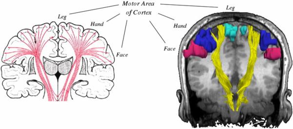
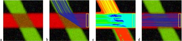
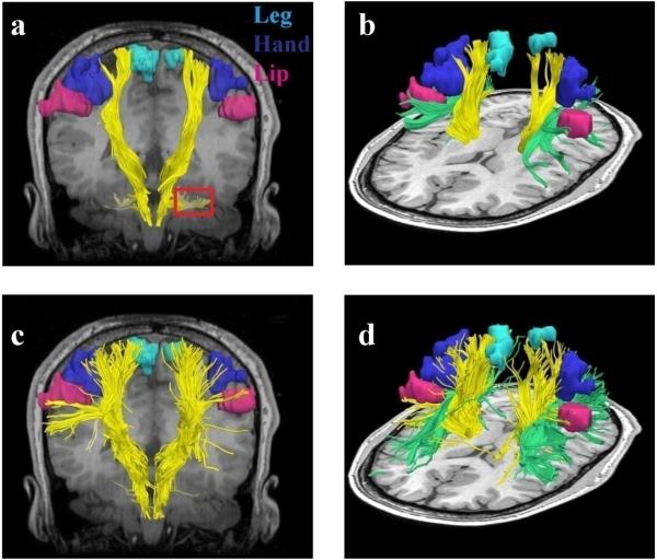
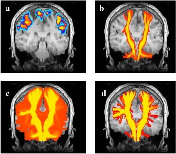
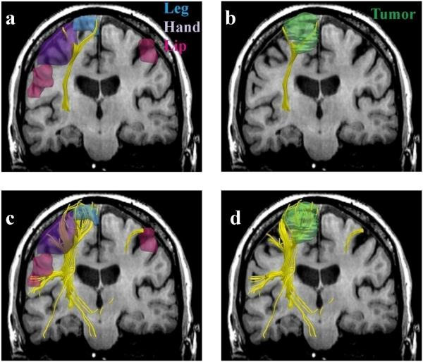
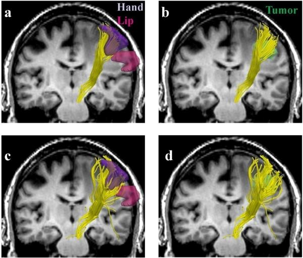
An inherent drawback of the traditional diffusion tensor model is its limited ability to provide detailed information about multidirectional fiber architecture within a voxel. This leads to erroneous fiber tractography results in locations where fiber bundles cross each other. This may lead to the inability to visualize clinically important tracts such as the lateral projections of the corticospinal tract. In this report, we present a deterministic two-tensor eXtended Streamline Tractography (XST) technique, which successfully traces through regions of crossing fibers. We evaluated the method on simulated and in vivo human brain data, comparing the results with the traditional single-tensor and with a probabilistic tractography technique. By tracing the corticospinal tract and correlating with fMRI-determined motor cortex in both healthy subjects and patients with brain tumors, we demonstrate that two-tensor deterministic streamline tractography can accurately identify fiber bundles consistent with anatomy and previously not detected by conventional single-tensor tractography. When compared to the dense connectivity maps generated by probabilistic tractography, the method is computationally efficient and generates discrete geometric pathways that are simple to visualize and clinically useful. Detection of crossing white matter pathways can improve neurosurgical visualization of functionally relevant white matter areas.


Figures

FIG. 1
FIG. 1
Anatomic relationship of corticospinal tracts as they connect from the internal capsule to the various motor regions of the cortex. Conventional single tensor tractography is only able to demonstrate the fibers from the motor tracts (shown in yellow) leading to the leg area of the motor cortex (turquoise) and cannot resolve those fibers leading to the hand (blue) or face (magenta) areas.
FIG. 2
FIG. 2
Simulated data. (a) The simulated 60° fiber crossing. Inside the brown region (middle), the two fibers are crossing each other. Outside the crossing region there is one anisotropic tensor per fiber and the direction is color-coded. (b) Single tensor tractography when seeded in the region bounded by the yellow box. (c) Connection map from Probabilistic tractography when seeded in the same region (voxels are color coded from 5000 (blue) to 4 (red) samples passing through the voxel). (d) Tractography based on XST.
FIG. 3
FIG. 3
Single tensor vs. XST when seeded in the internal capsule. Fibers from single tensor tractography and the fMRI activation areas are shown in (a) coronal and (b) lateral view. Note that where the CST passes through the pons, single tensor tractography demonstrates divergent fibers leading into the cerebellum (region marked in red). Fibers from the superior longitudinal fasciculus (SLF) (green) are also shown. (c) Fibers traced from XST are shown in (c) coronal and (d) lateral view. The fibers are able to reach all three fMRI activation areas. (d) shows areas of crossing between the SLF and CST.
FIG. 4
FIG. 4
Tracing the motor tract when seeded from the posterior limb of the internal capsule. To generate a comparable visualization to probabilistic tractography for all methods, a coronal maximum intensity projection (MIP) map is shown for all tractography and fMRI data, overlaid on a representative coronal anatomical image. (a) The FMRI activation areas. (b) Single tensor tractography (voxels are color coded based on the number of trajectories passing through them, followed by the MIP). (c) Probabilistic tractography. (d) two-tensor XST. In (b), (c), and (d) the colored regions show voxels containing at least one fiber trajectory.
FIG. 5
FIG. 5
Single (a,b) vs. two-tensor XST (c,d) tractography results on a 64-year old caucasian female with left frontoparietal meningioma. Fibers were seeded in the internal capsule and those intersecting fMRI activations were retained. Three-dimensional surface models represent (a, c) fMRI areas for the leg (blue), hand (purple) and the lip (pink) and (b, d) tumor (green).
FIG. 6
FIG. 6
Single (a,b) vs. two-tensor XST (c,d) tractography results on a 64-year old caucasian male with right metastatic melanoma. Fibers were seeded in the internal capsule and those intersecting fMRI activations were retained. Three-dimensional surface models represent (a, c) fMRI areas for the hand (purple) and the lip (pink) and (b, d) tumor (green).

References

    1. Akaike H. A new look at the statistical model identification. IEEE Transactions on Automatic Control. 1974;19:716–723.
    1. Alexander AL, Hasan KM, Lazar M, Tsuruda JS, Parker DL. Analysis of partial volume effects in diffusion-tensor MRI. Magnetic Resonance in Medicine. 2001;45:770–780. - PubMed
    1. Alexander DC. Multiple-Fiber Reconstruction Algorithms for Diffusion MRI. Annals of the New York Academy of Sciences. 2005;1046:113–133. - PubMed
    1. Alexander DC, Barker GJ, Arridge SR. Detection and modeling of non-Gaussian apparent diffusion coefficient profiles in human brain data. Magnetic Resonance in Medicine. 2002;48:331–340. - PubMed
    1. Basser PJ. Inferring microstructural features and the physiological state of tissues from diffusion-weighted images. NMR in Biomedicine. 1995;8:333–344. - PubMed

Publication types