. 2008 Sep 10;314(15):2884-94.
doi: 10.1016/j.yexcr.2008.06.003. Epub 2008 Jun 9.

The LIM protein LIMD1 influences osteoblast differentiation and function

Affiliations

The LIM protein LIMD1 influences osteoblast differentiation and function

Hilary F Luderer et al. Exp Cell Res. .

Abstract

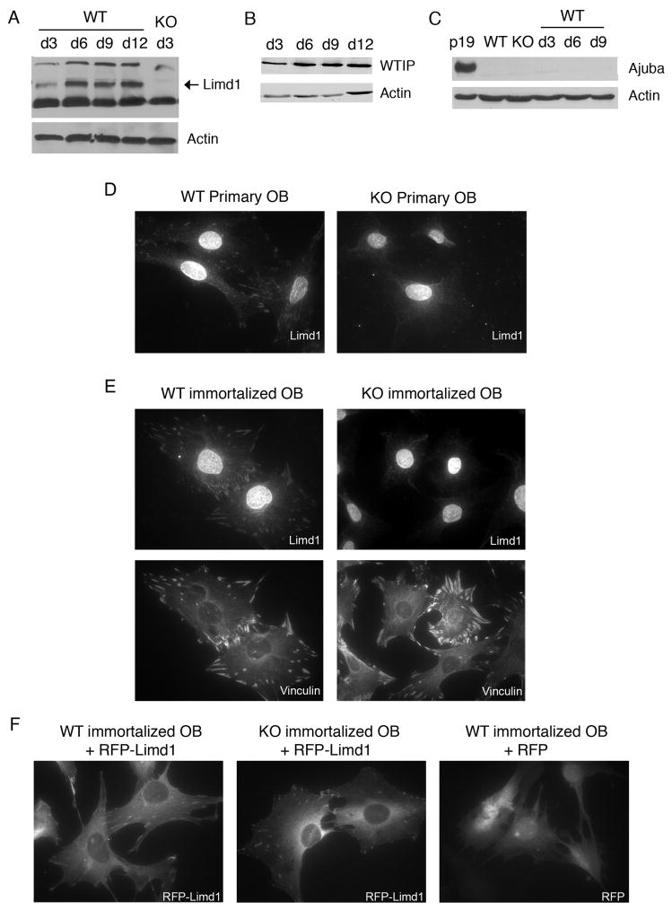
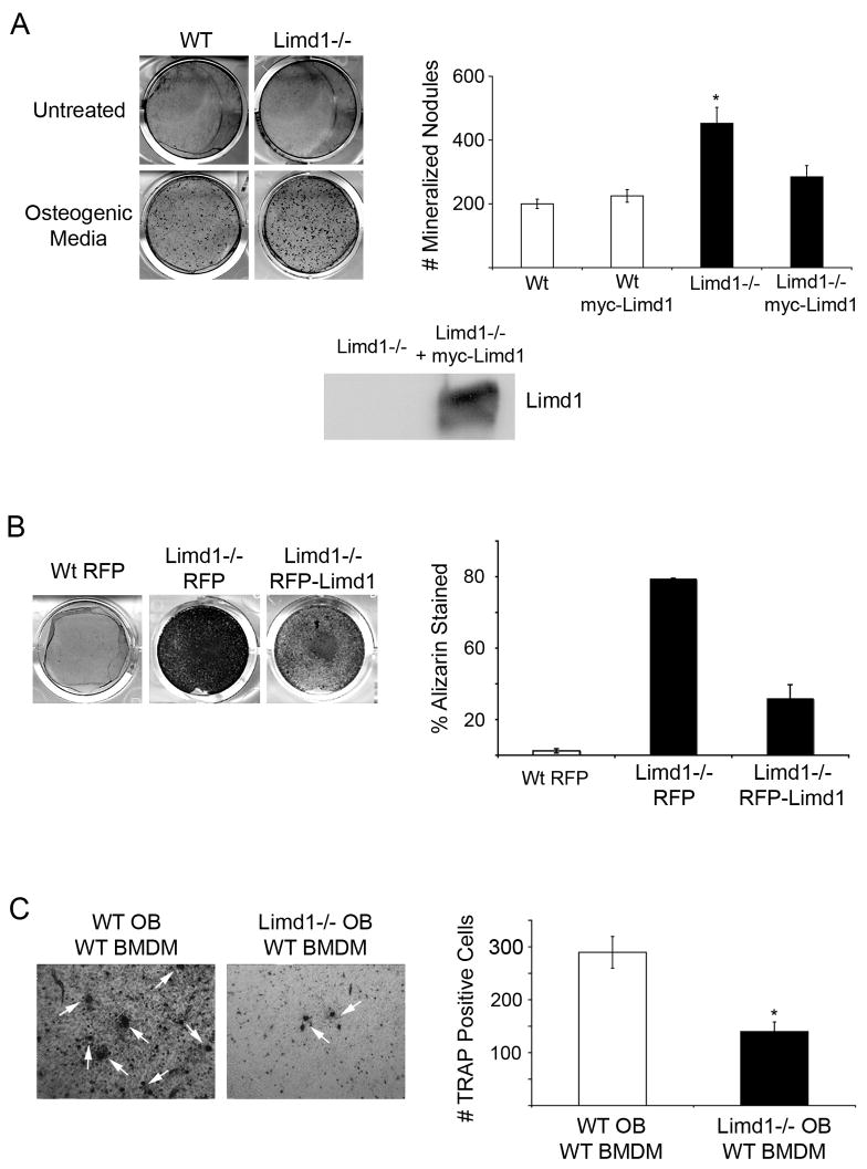
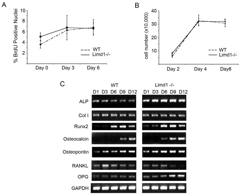
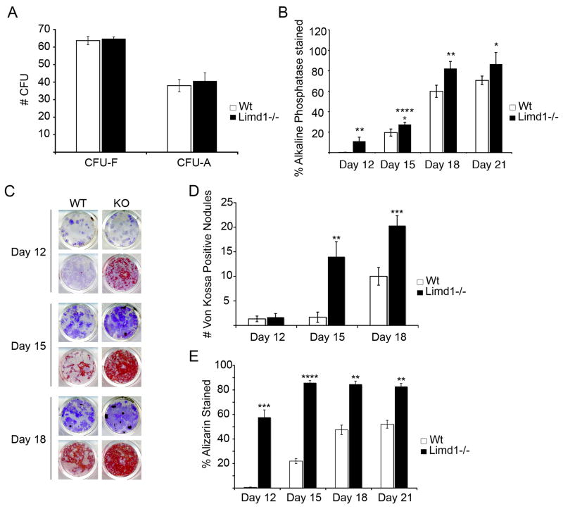
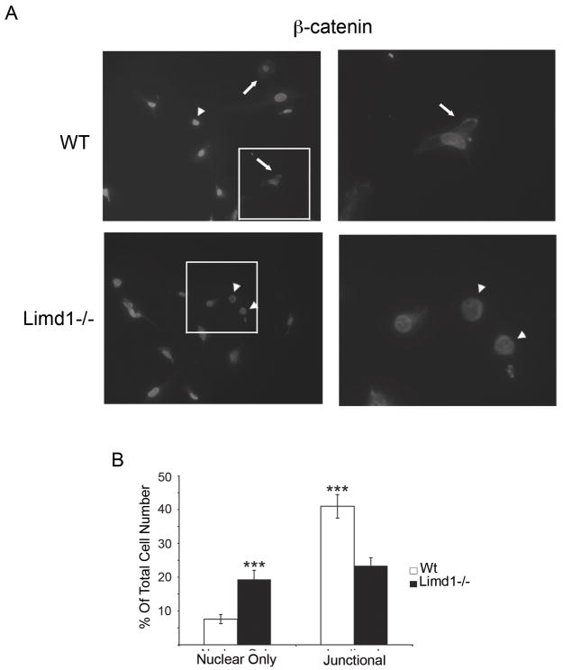
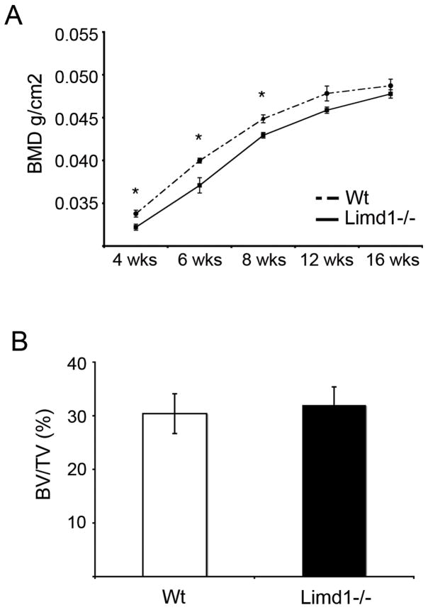
The balance between bone resorption and bone formation involves the coordinated activities of osteoblasts and osteoclasts. Communication between these two cell types is essential for maintenance of normal bone homeostasis; however, the mechanisms regulating this cross talk are not completely understood. Many factors that mediate differentiation and function of both osteoblasts and osteoclasts have been identified. The LIM protein Limd1 has been implicated in the regulation of stress osteoclastogenesis through an interaction with the p62/sequestosome protein. Here we show that Limd1 also influences osteoblast progenitor numbers, differentiation, and function. Limd1(-/-) calvarial osteoblasts display increased mineralization and accelerated differentiation. While no significant differences in osteoblast number or function were detected in vivo, bone marrow stromal cells isolated from Limd1(-/-) mice contain significantly more osteoblast progenitors compared to wild type controls when cultured ex vivo. Furthermore, we observed a significant increase in nuclear beta-catenin staining in differentiating Limd1(-/-) calvarial osteoblasts suggesting that Limd1 is a negative regulator of canonical Wnt signaling in osteoblasts. These results demonstrate that Limd1 influences not only stress osteoclastogenesis but also osteoblast function and osteoblast progenitor commitment. Together, these data identify Limd1 as a novel regulator of both bone osetoclast and bone osteoblast development and function.


Figures

Fig. 1
Fig. 1
Limd1 protein expression and localization in calvarial osteoblasts. (A–C) Primary calvarial osteoblasts were cultured in osteogenic conditions for 3, 6, 9, or 12 days, lysed, and equal amounts of protein run on SDS-PAGE then Western blotted for the presence of the Ajuba-LIM family members Limd1 (A), WTIP (B) and Ajuba (C). Actin was used as a loading control. Primary (D) or immortalized (E) Wt or Limd1−/− calvarial osteoblasts were fixed, permeabilized and stained with Limd1 or vinculin antisera. (F) Immortalized wt or Limd1−/−calvarial osteoblasts transduced with RFP or RFP-Limd1, as indicated.
Fig. 2
Fig. 2. Limd1−/− calvarial osteoblasts display increased mineralization and support less osteoclastogenesis than Wt controls
(A) Primary Wt or Limd1−/− calvarial osteoblasts were cultured in normal or osteogenic media for 12 days then fixed and stained with Alizarin Red S to detect mineralization. The number of Alizarin positive nodules were counted and graphed. Data is a compilation of three different experiments, all performed in triplicate. For rescue experiments, Limd1−/− calvarial osteoblasts were infected with either myc-empty or myc-Limd1 retroviruses before exposure to osteogenic culture conditions. Aliquots of Limd1−/− cells infected with myc-empty or myc-Limd1 were lysed and equal amounts of protein run on SDS-PAGE then Western blotted for the presence of Limd1. (B) Immortalized Wt or Limd1−/− calvarial osteoblast were cultured in osteogenic conditions for 12 days then fixed, stained with Alizarin Red S, and the percentage of Alizarin Red S staining determined and graphed. (C) Equal numbers of primary Wt or Limd1−/− calvarial osteoblasts and wt BMDM were co-cultured in the presence of vitamin D for 10 days then fixed and TRAP stained to identify osteoclasts (white arrows). The number of TRAP positive osteoclasts was quantified and graphed. For rescue experiments, Limd1−/− calvarial osteoblasts were infected with either myc-empty or myc-Limd1 retroviruses before co-culturing. *Indicates a significant difference between Limd1−/− and Wt (*p<0.05, **p<0.01, ****p<0.001, student’s paired t-test).
Fig. 3
Fig. 3
Limd1 influences osteoblast differentiation but not proliferation. (A) Primary wt or Limd1−/− calvarial osteoblasts were cultured in osteogenic media for 0, 3, or 6 days then incubated with BrdU, fixed and stained. The number of BrdU positive nuclei was normalized to the total number of nuclei. (B) Equal numbers of wt or Limd1−/− calvarial osteoblasts (100,000) were plated and cultured in osteogenic conditions for the time indicated. Total cell numbers were determined and graphed for comparison. (C) RT-PCR analysis of osteoblast-specific gene expression in RNA isolated from primary Wt or Limd1−/− calvarial osteoblasts cultured in osteogenic media for 1, 3, 6, 9, and 12 days. ALP – alkaline phosphatase; Col I – type 1 collagen; RANKL – receptor activator of NF-kB ligand; OPG – osteoprotegrin; GAPDH – glyceraldehydes-3-phosphate dehydrogenase
Fig. 4
Fig. 4
Limd1−/− mice have increased numbers of osteoblast progenitors. (A) A comparison of the number of hematoxylin positive fibroblast colony forming units (CFU-F) or Oil Red O positive adipocyte colony forming unit (CFU-A) present after culturing Limd1−/− and Wt mice bone marrow stromal cells in normal or adipogenic media for 9 days. (B) A comparison of the number of alkaline phosphatase positive CFU-O present after culturing Limd1−/− and wt bone marrow stromal cells in osteogenic media for the number of days indicated. (C–E) Limd1−/− and Wt marrow stromal cells were cultured in osteogenic media for the number of days indicated days then fixed and stained for the presence of alkaline phosphatase (purple, all wells). Mineralization was visualized by Von Kossa (black, top row for each time point) or Alizarin Red S (red, bottom row for each time point) and the number of Von Kossa positive CFU-O (D) or Alizarin positive CFU-O (E) enumerated. *Indicates a significant difference between Limd1−/− and Wt (*p<0.05, **p<0.01, ***p<0.005, ****p<0.001, as determined by the student’s paired t-test).
Fig. 5
Fig. 5
Limd1−/− calvarial cells have more nuclear β-catenin than Wt controls. (A) Primary Wt or Limd1−/− calvarial osteoblasts were fixed, permeabilized and stained for β-catenin and DAPI. Cells with nuclear only β-catenin (arrowheads) and junctional β-catenin (arrows) are highlighted. (B) The percentage of cells with nuclear only β-catenin and the percentage of cells containing junctional β-catenin was determined and graphed. (***p<0.005, as determined by the student’s paired t-test).
Fig. 6
Fig. 6
Basal bone density is not increased in Limd1−/− mice. (A) Dual energy x-ray absorptiometry (DEXA) analysis was performed on a cohort of Limd1−/− (n=9) and Wt (n=8) mice at 4, 6, 8 12 and 16 weeks of age. (B) Micro-CT analysis of tibiae from Limd1−/− (n=3) and Wt (n=3) mice at 6 weeks of age. (*p<0.05, as determined by the student’s paired t-test).

Similar articles

Cited by

References

    1. Roodman GD. Regulation of osteoclast differentiation. Ann N Y Acad Sci. 2006;1068:100–9. - PubMed
    1. Rogers A, Eastell R. Circulating osteoprotegerin and receptor activator for nuclear factor kappaB ligand: clinical utility in metabolic bone disease assessment. J Clin Endocrinol Metab. 2005;90:6323–31. - PubMed
    1. Kiss H, Kedra D, Yang Y, Kost-Alimova M, Kiss C, O’Brien KP, Fransson I, Klein G, Imreh S, Dumanski JP. A novel gene containing LIM domains (LIMD1) is located within the common eliminated region 1 (C3CER1) in 3p21.3. Hum Genet. 1999;105:552–9. - PubMed
    1. Crawford AW, Beckerle MC. Purification and characterization of zyxin, an 82,000-dalton component of adherens junctions. J Biol Chem. 1991;266:5847–53. - PubMed
    1. Petit MM, Mols R, Schoenmakers EF, Mandahl N, Van de Ven WJ. LPP, the preferred fusion partner gene of HMGIC in lipomas, is a novel member of the LIM protein gene family. Genomics. 1996;36:118–29. - PubMed

Publication types