

Ultrahigh-speed optical coherence tomography for three-dimensional and en face imaging of the retina and optic nerve head
- PMID: 18658089
- PMCID: PMC2743183
- DOI: 10.1167/iovs.08-2127
Ultrahigh-speed optical coherence tomography for three-dimensional and en face imaging of the retina and optic nerve head
Abstract
Purpose: To demonstrate ultrahigh-speed optical coherence tomography (OCT) imaging of the retina and optic nerve head at 249,000 axial scans per second and a wavelength of 1060 nm. To investigate methods for visualization of the retina, choroid, and optic nerve using high-density sampling enabled by improved imaging speed.
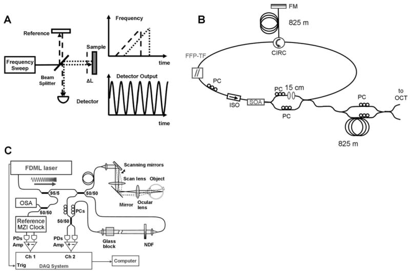
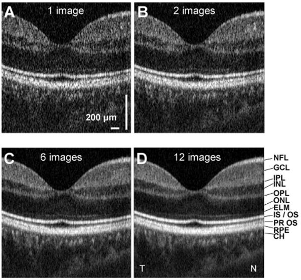
Methods: A swept-source OCT retinal imaging system operating at a speed of 249,000 axial scans per second was developed. Imaging of the retina, choroid, and optic nerve were performed. Display methods such as speckle reduction, slicing along arbitrary planes, en face visualization of reflectance from specific retinal layers, and image compounding were investigated.
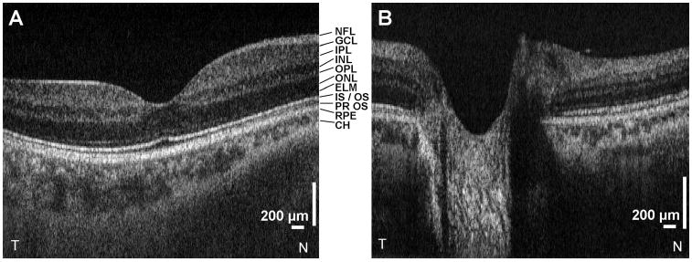
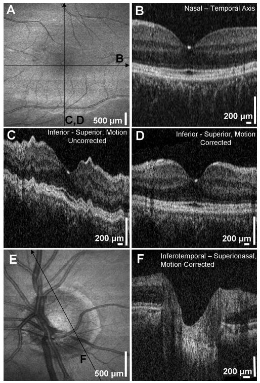
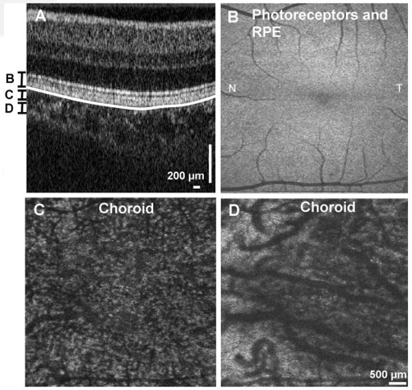
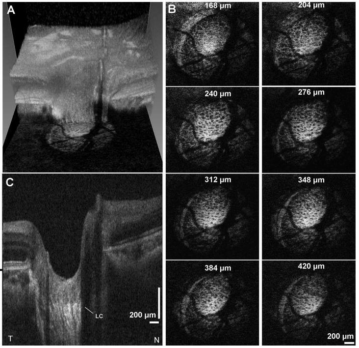
Results: High-definition and three-dimensional (3D) imaging of the normal retina and optic nerve head were performed. Increased light penetration at 1060 nm enabled improved visualization of the choroid, lamina cribrosa, and sclera. OCT fundus images and 3D visualizations were generated with higher pixel density and less motion artifacts than standard spectral/Fourier domain OCT. En face images enabled visualization of the porous structure of the lamina cribrosa, nerve fiber layer, choroid, photoreceptors, RPE, and capillaries of the inner retina.
Conclusions: Ultrahigh-speed OCT imaging of the retina and optic nerve head at 249,000 axial scans per second is possible. The improvement of approximately 5 to 10x in imaging speed over commercial spectral/Fourier domain OCT technology enables higher density raster scan protocols and improved performance of en face visualization methods. The combination of the longer wavelength and ultrahigh imaging speed enables excellent visualization of the choroid, sclera, and lamina cribrosa.
Conflict of interest statement
Disclosure:
Figures

References
-
- Puliafito CA, Hee MR, Lin CP, et al. Imaging of macular diseases with optical coherence tomography. Ophthalmology. 1995;102(2):217–229. - PubMed
-
- Schuman JS, Puliafito CA, Fujimoto JG. Optical Coherence Tomography of Ocular Diseases. 2nd. Thorofare, NJ: Slack Inc.; 2004.
-
- Fercher AF, Hitzenberger CK, Kamp G, Elzaiat SY. Measurement of intraocular distances by backscattering spectral interferometry. Opt Commun. 1995;117(1–2):43–48.
-
- Häusler G, Lindner MW. “Coherence radar” and “spectral radar”: new tools for dermatological diagnosis. J Biomed Opt. 1998;3(1):21–31. - PubMed
Publication types
MeSH terms
Grants and funding
LinkOut - more resources
Full Text Sources
Other Literature Sources
Research Materials
