Comparative Study
. 2008 Nov;49(11):4837-43.
doi: 10.1167/iovs.08-2115. Epub 2008 Jul 24.

DNA cross-linking, double-strand breaks, and apoptosis in corneal endothelial cells after a single exposure to mitomycin C

Affiliations
Comparative Study

DNA cross-linking, double-strand breaks, and apoptosis in corneal endothelial cells after a single exposure to mitomycin C

Danny S Roh et al. Invest Ophthalmol Vis Sci. 2008 Nov.

Abstract

Purpose: To investigate the cellular effects of mitomycin C (MMC) treatment on corneal endothelial (CE) cells at clinically relevant applications and dosages.

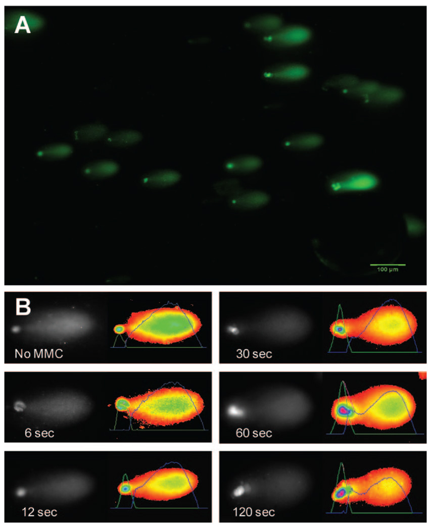
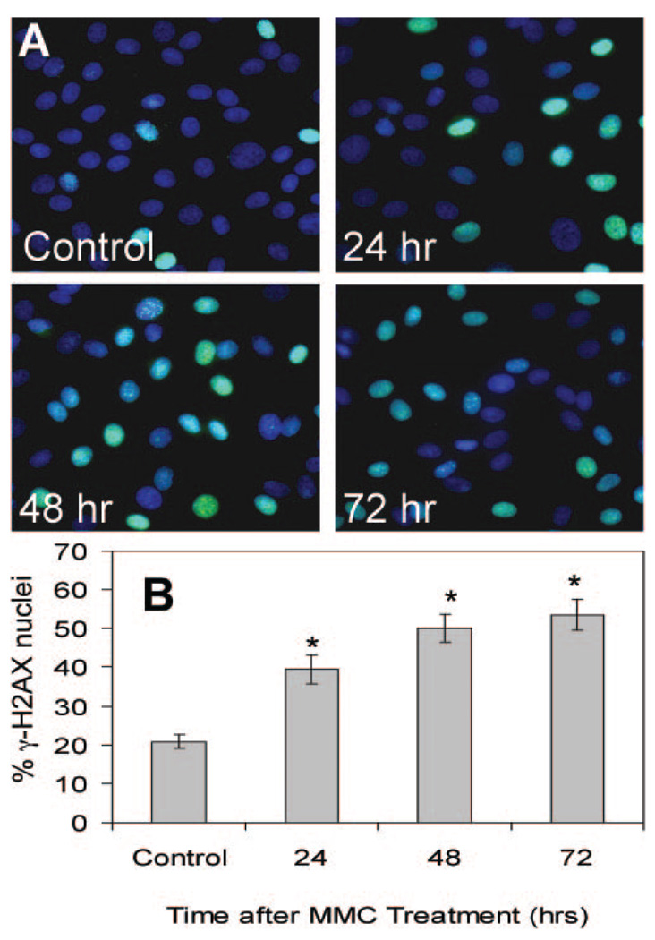
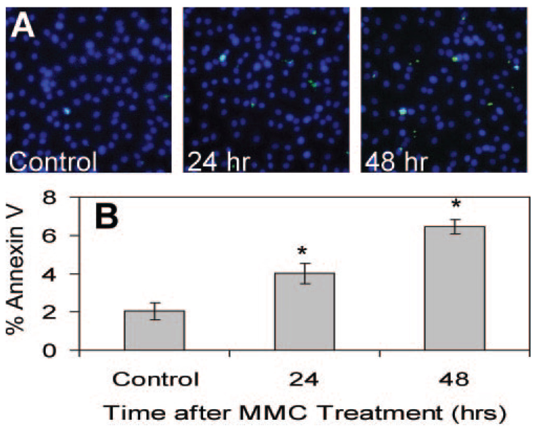
Methods: Radial and posterior diffusion of MMC was determined by an Escherichia coli growth inhibition bioassay. A modified version of the comet assay (single cell gel electrophoresis) was used to detect DNA cross-linking. Immunostaining detected the nuclear phosphorylated histone variant H2AX (gamma-H2AX) indicating DNA double-strand breaks. Apoptosis in MMC-treated cells was detected with annexin V staining.

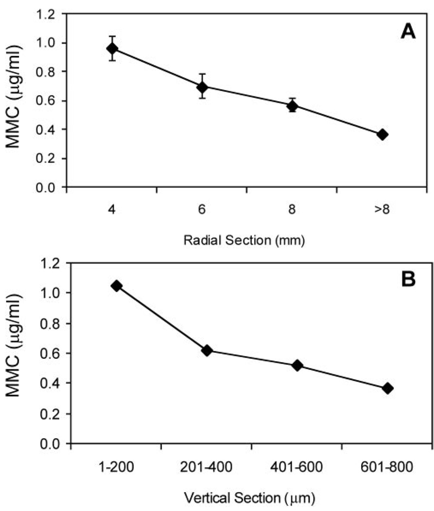
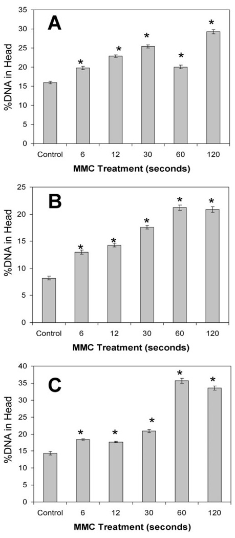
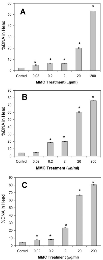
Results: Topical application of 0.02% MMC to intact goat globes resulted in MMC in the CE at 0.37 microg/mL and produced a significant increase in CE DNA cross-linking with as little as 6 seconds of topical MMC treatment. DNA cross-linking was also demonstrated in cultured CE cells by using MMC exposures similar to those detected in CE of intact eyes. Such MMC treatment of CE produced elevated and persistent gamma-H2AX-positive cells indicative of DNA double-strand breaks. Similarly, there was an increase in the proportion of apoptotic CE cells, evidenced by positive annexin V staining.

Conclusions: The results demonstrate that exposure to MMC at times and concentrations commonly used in refractive surgery produces cross-linking of corneal endothelial DNA, persistent DNA damage, and endothelial death via apoptosis. Current practices of MMC application during refractive surgeries may increase the potential for long-term and permanent deleterious effects on the health of the corneal endothelium.


Figures

FIGURE 1
FIGURE 1
Corneal concentration of MMC 2 minutes after a single topical administration of 0.02% MMC on a whole globe, as determined by growth inhibition bioassay. The zone of inhibition of E. coli growth was compared to a standard curve to determine MMC concentrations. (A) MMC concentration decreases radially, away from the site of application. (B) CE concentration is approximately 0.37 µg/mL at the depth of the endothelium (601–800 µm). Mean ± SEM.
FIGURE 2
FIGURE 2
Single cell gel electrophoresis (modified alkaline comet assay) of CE cell DNA. (A) Representative images of individual CE cell DNA after electrophoresis show migration patterns with a comet-like appearance. (B) Individual comets are oriented with the head to the left. The image on the right of each shows a pseudocolor of the image with an overlaid graphic representation of staining intensity. This intensity is proportional to the amount of DNA at that location in the gel. Area under these curves was used calculate %DNA-in-head. With increasing length of treatment of MMC, more DNA was observed in the head of comets.
FIGURE 3
FIGURE 3
CE DNA cross-linking induced by MMC treatment of whole globes. %DNA-in-head was calculated for comets (as in Fig. 2) derived from CE cells after topical MMC treatment of intact goat eyes. (A) 0.02% MMC treatment followed epithelial debridement. (B) MMC treatment followed epithelial debridement and 75 µm stromal ablation. (C) Eyes treated with MMC after epithelial debridement (as in A) were incubated 24 hours at 37°C, to allow DNA repair. *Significant difference from control; ANOVA P < 0.0001, Dunnett P < 0.01. Mean ± SEM.
FIGURE 4
FIGURE 4
Sensitivity of CE cells to DNA cross-linking by MMC. Goat CE cells in culture were treated with a range of MMC concentrations, and DNA cross-linking was determined using the comet assay as described in Figure 2 at three exposure times. (A) Ten-minute exposure to MMC at all concentrations induced significant DNA cross-links compared to controls. (B) Thirty-minute exposure to MMC at all concentrations (except 0.02 µg/mL, Dunnett P > 0.05) induced significant DNA cross-links compared with controls. (C) Sixty-minute exposure to MMC at all concentrations induced significant DNA cross-links compared with the control. *Significant differences from control; ANOVA P < 0.0001, Dunnett P < 0.01. Mean ± SEM.
FIGURE 5
FIGURE 5
H2AX phosphorylation (γ-H2AX) induced by MMC treatment. (A) Confluent CE cells were treated with MMC at 0.4 µg/mL for 30 minutes and nuclei were stained with DAPI (blue) and anti-γ-H2AX anti-body (green) at 24, 48, and 72 hours after treatment. (B) The proportion of nuclei with γ-H2AX staining was similar to that in (A). *Significant differences from control; ANOVA P < 0.0001, Dunnett P < 0.01. The control cells were assayed at 72 hours. Mean ± SEM.
FIGURE 6
FIGURE 6
MMC exposure initiates endothelial apoptosis. (A) Confluent CE cells were treated with MMC 0.4 µg/mL for 30 minutes and stained with Hoechst 33342 (blue) and annexin V-FITC (green) 24 and 48 hours later. Cells positive for annexin V-FITC are apoptotic. (B) The proportion of apoptotic cells after MMC treatment was significantly elevated above the untreated control. *Significant differences from control; ANOVA P < 0.0001, Dunnett P < 0.01. The control cells were assayed at 48 hours. Mean ± SEM.

References

    1. Chabner BA, Amrein PC, Druker BJ, et al. Antineoplastic agents. In: Brunton LL, Lazo JS, Parker KL, editors. Goodman and Gilman’s The Pharmacological Basis of Therapeutics. 11th ed. New York: McGraw Hill; 2006.
    1. Abraham LM, Selva D, Casson R, Leibovitch I. Mitomycin: clinical applications in ophthalmic practice. Drugs. 2006;66:321–340. - PubMed
    1. Majmudar PA, Forstot SL, Dennis RF, et al. Topical mitomycin-C for subepithelial fibrosis after refractive corneal surgery. Ophthalmology. 2000;107:89–94. - PubMed
    1. Carones F, Vigo L, Scandola E, Vacchini L. Evaluation of the prophylactic use of mitomycin-C to inhibit haze formation after photorefractive keratectomy. J Cataract Refract Surg. 2002;28:2088–2095. - PubMed
    1. Lane HA, Swale JA, Majmudar PA. Prophylactic use of mitomycin-C in the management of a buttonholed LASIK flap. J Cataract Refract Surg. 2003;29:390–392. - PubMed

Publication types

MeSH terms