Comparative Study
. 2008 Oct 15;178(8):822-31.
doi: 10.1164/rccm.200712-1894OC. Epub 2008 Jul 24.

Potential role of high-mobility group box 1 in cystic fibrosis airway disease

Affiliations
Comparative Study

Potential role of high-mobility group box 1 in cystic fibrosis airway disease

Steven M Rowe et al. Am J Respir Crit Care Med. .

Abstract

Rationale: High-mobility group box 1 (HMGB1) is a potent inflammatory mediator elevated in sepsis and rheumatoid arthritis, although its role in cystic fibrosis (CF) lung disease is unknown.

Objectives: To determine whether HMGB1 contributes to CF lung inflammation, including neutrophil chemotaxis and lung matrix degradation.

Methods: We used sputum and serum from subjects with CF and a Scnn1b-transgenic (Scnn1b-Tg) mouse model that overexpresses beta-epithelial Na(+) channel in airways and mimics the CF phenotype, including lung inflammation. Human secretions and murine bronchoalveolar lavage fluid (BALF) was assayed for HMGB1 by Western blot and ELISA. Neutrophil chemotaxis was measured in vitro after incubation with human neutrophils. The collagen fragment proline-glycine-proline (PGP) was measured by tandem mass spectroscopy.

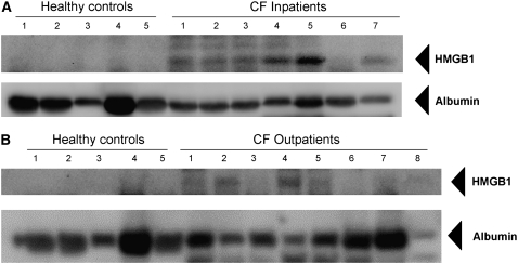
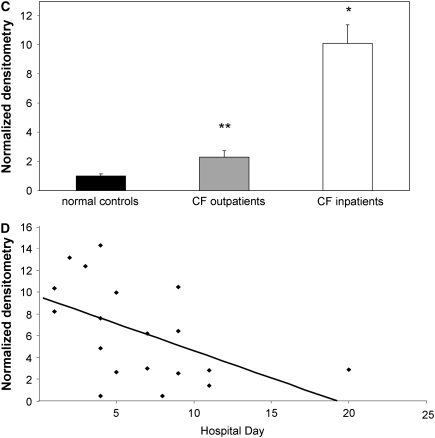
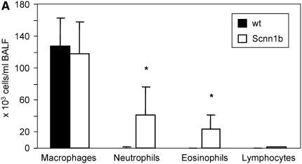
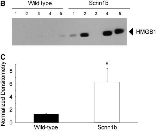
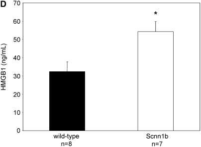
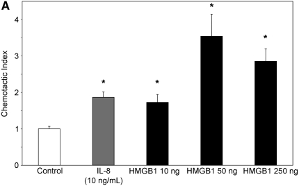
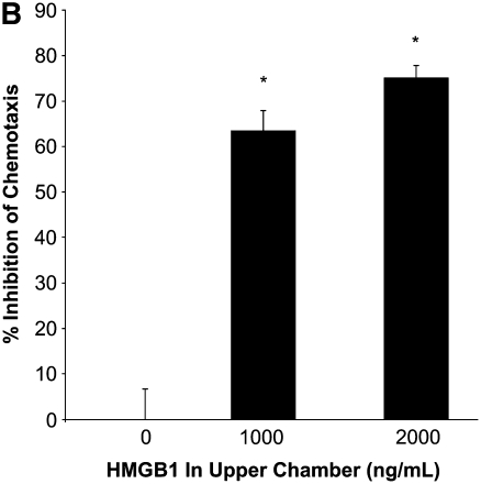
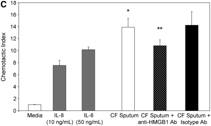
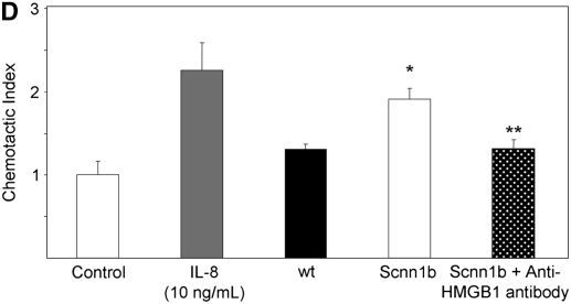
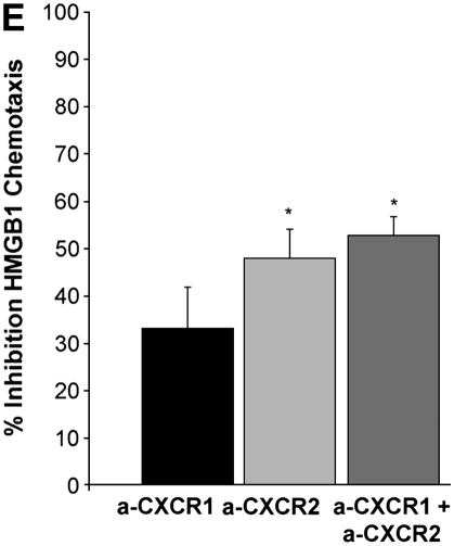
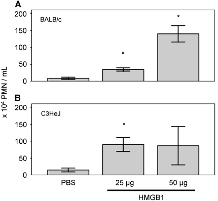
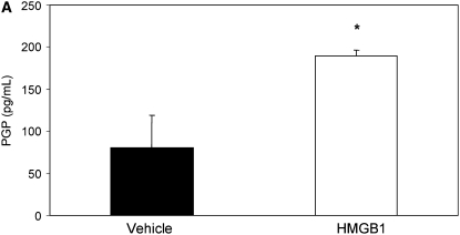
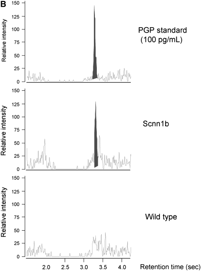
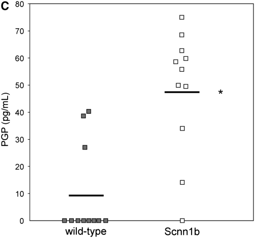
Measurements and main results: HMGB1 was detected in CF sputum at higher levels than secretions from normal individuals. Scnn1b-Tg mice had elevated levels of HMGB1 by Western blot and ELISA. We demonstrated that dose-dependent chemotaxis of human neutrophils stimulated by purified HMGB1 was partially dependent on CXC chemokine receptors and that this could be duplicated in CF sputum and BALF from Scnn1b-Tg mice. Neutralization by anti-HMGB1 antibody, in both the sputum and BALF-reduced chemotaxis, which suggested that HMGB1 contributed to the chemotactic properties of these samples. Intratracheal administration of purified HMGB1 induced neutrophil influx into the airways of mice and promoted the release of PGP. PGP was also elevated in Scnn1b-Tg mice and CF serum.

Conclusions: HMGB1 expression contributes to pulmonary inflammation and lung matrix degradation in CF airway disease and deserves further investigation as a biomarker and potential therapeutic target.


Figures

<b>Figure 1.</b>
Figure 1.
High-mobility group box 1 (HMGB1) levels are elevated in sputum from subjects with cystic fibrosis (CF) and inversely correlated with the duration of intravenous antimicrobial therapy. HMGB1 detected by Western blot in spontaneously expectorated sputum from (A) subjects with CF hospitalized for CF exacerbation and (B) outpatients with CF was significantly greater than airway secretions from normal subjects. HMGB1 has a calculated molecular weight of 24.9 kD and migrates as a 29-kD band under sodium dodecyl sulfate–polyacrylamide gel electrophoresis conditions. Albumin loading control is shown for comparison. (C) Summary data of densitometry results showing that HMGB1 levels were significantly elevated in sputa from patients with CF than in secretions from healthy control subjects. All specimens were standardized for total protein concentration and compared by relative densitometry; *P < 0.001, **P < 0.05; n = 5 controls, 23 inpatients with CF, and 9 outpatients with CF, ±SEM. (D) HMGB1 levels in sputum from patients with CF quantified by Western blot densitometry were inversely correlated with the number of days subject was hospitalized for intravenous antimicrobial therapy, reflecting reduced levels of HMGB1 after systemic antibiotic therapy; r = −0.51, P < 0.05.
<b>Figure 2.</b>
Figure 2.
High-mobility group box 1 (HMGB1) levels are elevated in the lungs of Scnn1b-transgenic (Scnn1b-Tg) mice. (A) Neutrophils and eosinophils are significantly increased in bronchoalveolar lavage fluid (BALF) samples obtained from Scnn1b-Tg mice compared with wild-type littermate controls. BALF samples were procured in a paired fashion at 5 weeks of life; P < 0.005, n = 15 per genotype. (B) HMGB1 was detected after loading 30 μl BALF aliquots and analyzed by Western blot using sodium dodecyl sulfate–polyacrylamide gel electrophoresis (SDS-PAGE) and immunoblot with monoclonal antibody directed against HMGB1. HMGB1 has a calculated molecular weight of 24.9 kD and migrates as a 29-kD band under SDS-PAGE conditions. (C) Summary of data representing normalized densitometry of wild-type versus Scnn1b-Tg mice; *P < 0.05 by densitometry analysis, n = 10 per genotype. (D) HMGB1 was detected by ELISA; *P < 0.05, n = 8 wild-type and 7 Scnn1b-Tg mice. wt = wild type.
<b>Figure 3.</b>
Figure 3.
High-mobility group box 1 (HMGB1) is a potent chemoattractant for isolated human neutrophils in vitro, contributes to polymorphonuclear leukocyte chemotaxis elicited by sputum from subjects with cystic fibrosis (CF) and from bronchoalveolar lavage fluid (BALF) of Scnn1b-transgenic (Scnn1b-Tg) mice, and can be partially inhibited by neutralizing antibody. (A) Chemotaxis of various concentrations of recombinant human HMGB1 was compared with medium (negative control) and IL-8 (positive control). Migration was measured by quantitative photomicrograph after 1 hour incubation and standardized to cell count found in medium control; *P < 0.005 versus medium control, n = 5, ±SEM. (B) HMGB1 is chemotactic rather than chemokinetic, as evidenced by inhibition of chemotaxis toward HMGB1 in the lower chamber (2,000 ng/ml) by placement of HMGB1 in the upper chamber; *P < 0.05 versus control, n = 3 per condition, ±SEM. (C) CF sputum was evaluated alone, with anti-HMGB1 neutralizing antibody or isotype control antibody. Medium and IL-8 positive control are provided for comparison; *P < 0.001 versus medium; **P < 0.01 versus CF sputum; n = 6, ±SEM. (D) BALF from Scnn1b-Tg mice was compared with wild-type littermate controls with and without the presence of anti-HMGB1 neutralizing antibody; *P < 0.01 versus control and **P < 0.05 versus Scnn1b-Tg mice, n = 5, ±SEM. (E) HMGB1 induces human neutrophil chemotaxis by a CXCR2-dependent mechanism, as evidenced by blockade of HMGB1 (2,000 ng/ml)-induced chemotaxis after preincubation of isolated human neutrophils with anti-CXCR2 antibody. Neutralization with both anti-CXCR1 and anti-CXCR2 antibodies also inhibited chemotaxis. Anti-CXCR antibodies (25 μg/ml) were preincubated with neutrophils after isolation for 45 minutes at 4°C, as described in Methods; *P < 0.05, n = 3 per condition, ±SEM. wt = wild type.
<b>Figure 4.</b>
Figure 4.
Intratracheal injection of recombinant human high-mobility group box 1 (HMGB1) induces dose-dependent influx of neutrophils in bronchoalveolar lavage fluid. HMGB1 neutrophil chemotactic activity was observed in (A) wild-type BALB/c mice or (B) endotoxin-resistant C3H/HeJ mice. Mice were killed 24 hours after HMGB1 instillation; *P < 0.05, n = 3–6 per condition, ±SEM. PMN = polymorphonuclear leukocytes.
<b>Figure 5.</b>
Figure 5.
Intratracheal instillation of high-mobility group box 1 (HMGB1) triggers matrix collagen degradation and release of proline-glycine-proline (PGP) peptide, which is also found to be elevated in Scnn1b-transgenic (Scnn1b-Tg) mice bronchoalveolar lavage fluid (BALF) and in the serum of patients with cystic fibrosis (CF). (A) Instillation of HMGB1 at dosage sufficient to induce airway polymorphonuclear leukocyte influx also induces production of the collagen fragment PGP. Recombinant human HMGB1 (25 μg) was instilled in C3H/HeJ mice, which were then killed 24 hours later for BALF; *P < 0.05, n = 3 per condition, ±SEM. (B) Using electrospray ionization–liquid chromatography followed by tandem mass spectrometry, PGP was detected as a peak with retention time at 3.1 seconds and molecular weight splits of 270/70 and 270/116 (270/70 split shown in top panel; bold peak represents PGP standard [100 pg/ml]). The same peak was observed in BALF of Scnn1b-Tg mice (middle panel, representative section) but not in the BALF from wild-type littermate controls (bottom panel). (C) PGP was detected by electrospray ionization–liquid chromatography followed by tandem mass spectrometry and quantified by the relative standard curve method. BALF samples were obtained in a paired fashion from 5-week-old, sex-matched mice; *P < 0.005, n = 11 per genotype. (D) Serum samples obtained from normal nonsmoking individuals and subjects with CF hospitalized for acute pulmonary exacerbation were analyzed for PGP. Results were normalized to the expression seen in normal subjects; *P < 0.02, n = 10 normal subjects, 14 subjects with CF, ±SEM.

Similar articles

Cited by

References

    1. Wang H, Bloom O, Zhang M, Vishnubhakat JM, Ombrellino M, Che J, Frazier A, Yang H, Ivanova S, Borovikova L, et al. HMG-1 as a late mediator of endotoxin lethality in mice. Science 1999;285:248–251. - PubMed
    1. Abraham E, Arcaroli J, Carmody A, Wang H, Tracey KJ. HMG-1 as a mediator of acute lung inflammation. J Immunol 2000;165:2950–2954. - PubMed
    1. Kim JY, Park JS, Strassheim D, Douglas I, Diaz del Valle F, Asehnoune K, Mitra S, Kwak SH, Yamada S, Maruyama I, et al. HMGB1 contributes to the development of acute lung injury after hemorrhage. Am J Physiol Lung Cell Mol Physiol 2005;288:L958–L965. - PubMed
    1. Gardella S, Andrei C, Ferrera D, Lotti LV, Torrisi MR, Bianchi ME, Rubartelli A. The nuclear protein HMGB1 is secreted by monocytes via a non-classical, vesicle-mediated secretory pathway. EMBO Rep 2002;3:995–1001. - PMC - PubMed
    1. Bonaldi T, Talamo F, Scaffidi P, Ferrera D, Porto A, Bachi A, Rubartelli A, Agresti A, Bianchi ME. Monocytic cells hyperacetylate chromatin protein HMGB1 to redirect it towards secretion. EMBO J 2003;22:5551–5560. - PMC - PubMed

Publication types