. 2008 Oct 17;283(42):28660-9.
doi: 10.1074/jbc.M802645200. Epub 2008 Jul 24.

Increased dosage of Dyrk1A alters alternative splicing factor (ASF)-regulated alternative splicing of tau in Down syndrome

Affiliations

Increased dosage of Dyrk1A alters alternative splicing factor (ASF)-regulated alternative splicing of tau in Down syndrome

Jianhua Shi et al. J Biol Chem. .

Abstract

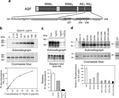
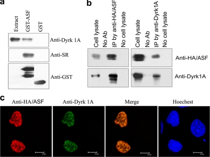
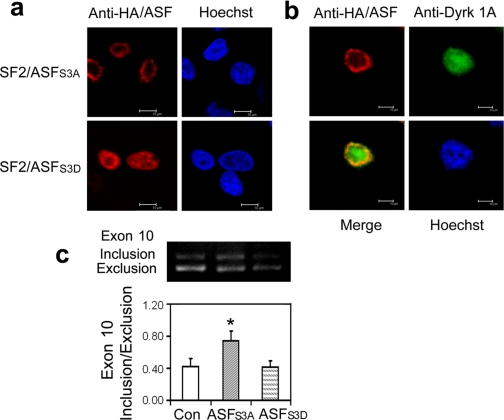
Two groups of tau, 3R- and 4R-tau, are generated by alternative splicing of tau exon 10. Normal adult human brain expresses equal levels of them. Disruption of the physiological balance is a common feature of several tauopathies. Very early in their life, individuals with Down syndrome (DS) develop Alzheimer-type tau pathology, the molecular basis for which is not fully understood. Here, we demonstrate that Dyrk1A, a kinase encoded by a gene in the DS critical region, phosphorylates alternative splicing factor (ASF) at Ser-227, Ser-234, and Ser-238, driving it into nuclear speckles and preventing it from facilitating tau exon 10 inclusion. The increased dosage of Dyrk1A in DS brain due to trisomy of chromosome 21 correlates to an increase in 3R-tau level, which on abnormal hyperphosphorylation and aggregation of tau results in neurofibrillary degeneration. Imbalance of 3R- and 4R-tau in DS brain by Dyrk1A-induced dysregulation of alternative splicing factor-mediated alternative splicing of tau exon 10 represents a novel mechanism of neurofibrillary degeneration and may help explain early onset tauopathy in individuals with DS.


Figures

FIGURE 1.
FIGURE 1.
Overexpression of ASF promotes tau exon 10 inclusion. a, overexpression of SR proteins affected tau E10 splicing. the pCI-SI9/LI10 mini-tau-gene was co-transfected with the same amount of pCEP4-SR proteins into HEK239-T cells. Total RNA was subjected to RT-PCR for measurement of tau exon 10 splicing after a 48-h transfection. b, expression of ASF promoted tau exon 10 inclusion. pCI-SI9/LI10 was co-transfected with pCEP4-ASF into various cell lines indicated under each panel. Tau exon 10 splicing was measured by RT-PCR after a 48-h transfection. c, knock-down of ASF by siRNA inhibited tau exon 10 inclusion. pCI-SI9/LI10 was co-transfected with ASF siRNA or scramble siRNA into HEK-293T cells. The expression of ASF was measured by Western blot (upper panel), and the tau exon 10 splicing was measured by RT-PCR after a 48-h transfection (lower panel). Con, control.
FIGURE 2.
FIGURE 2.
Dyrk1A phosphorylates ASF in vitro at Ser-227, Ser-234, and Ser-238. a, schematic of ASF domain structures and the serine residues (227, 234, and 238) in three conserved motifs (gray box) of Dyrk1A phosphorylation sites that were identified in this study. b, autoradiography of ASF phosphorylation by Dyrk1A in vitro. Recombinant GST-ASF was incubated with various concentrations of Dyrk1A indicated above each lane for 30 min at 30 °C and separated by SDS-PAGE and visualized with Coomassie Blue staining (middle panel). The last lane is Dyrk1A alone, without GST-ASF. After drying the gel, the 32P incorporated into ASF was measured by using a phosphorimaging device (BAS-1500, Fuji) (upper panel). The level of 32P was normalized by the protein level detected by Coomassie Blue staining was plotted to Dyrk1A concentration (lower panel). c, inhibition of Dyrk1A by EGCG decreased the phosphorylation of ASF in cultured cells. COS7 cells were transfected with pCEP4-ASF-HA for 45 h and then treated with 10 μm Tg003 and 50 μm EGCG to inhibit Clk and Dyrk1A, respectively. At the same time, [32P]orthophosphate was added to label the phosphoproteins. After a 3-h treatment and phospho-labeling, the cells were harvested, and the cell lysates were subjected to immunoprecipitation with anti-HA. The immunoprecipitated ASF-HA was analyzed by autoradiography and Western blot with anti-HA. The 32P incorporated into ASF-HA was normalized by ASF-HA level detected with anti-HA. Con, control; Pi, phosphate group. d, mutation of Ser to Ala at Ser-227, Ser-234, and/or Ser-238 inhibited ASF phosphorylation by Dyrk1A. Mutants of GST-ASF at the Ser residue indicated above each lane were phosphorylated by Dyrk1A (10 μg/ml) in vitro for 30 min. 32P incorporation into GST-ASFs was measured by using phosphorimaging analysis after the phosphorylation products were separated by SDS-PAGE. The 32P incorporated was normalized by protein level detected with Coomassie Blue staining. WT, wild type.
FIGURE 3.
FIGURE 3.
ASF interacts with Dyrk1A. a, Dyrk1A was pulled down from rat brain extract by GST-ASF. GST-ASF or GST coupled onto glutathione-Sepharose was incubated with rat brain extract. After washing, bound proteins were subjected to Western blots by using anti-GST, anti-SR protein, and anti-Dyrk1A. Only GST-ASF, but not GST, pulled down Dyrk1A. All these lanes are from the same blot from which unrelated lanes between the second and third lanes were removed. b, ASF and Dyrk1A could be co-immunoprecipitated by each other's antibodies. ASF tagged with HA and Dyrk1A were co-expressed in HEK-293T cells for 48 h. The cell extract was incubated with anti-HA or anti-Dyrk1A, and then protein G beads were added into the mixture. The bound proteins were subjected to Western blots by using antibodies indicated at the right of each blot. Dyrk1A and HA-ASF were co-immunoprecipitated by each other's antibodies, respectively. No Ab, no antibody; IP, immunoprecipitation. c, co-localization of ASF with Dyrk1A in nucleus. HA-ASF and Dyrk1A were co-transfected into HeLa cells. After a 48-h transfection, the cells were fixed and immunostained by anti-HA or anti-Dyrk1A and followed by TRITC-anti-rabbit IgG or FITC-anti-mouse IgG. Hoechst was used for nucleu staining.
FIGURE 4.
FIGURE 4.
Dyrk1A inhibits the role of ASF in tau exon 10 inclusion. a, overexpression of Dyrk1A inhibited the role of ASF in tau exon 10 inclusion. Mini-tau gene pCI-SI9/LI10 was co-transfected with ASF or Dyrk1A into COS7 cells for 48 h, and the total RNA was extracted and subjected for measurement of tau exon 10 splicing by using RT-PCR. Con, control. b, knock down of Dyrk1A by its siRNA elevated exon 10 inclusion induced by ASF. Mini-tau gene was co-transfected into HEK-293T cells with ASF and siRNA of Dyrk1A or its scrambled form, and then tau exon 10 splicing was analyzed by RT-PCR after a 48-h transfection. Expression of endogenous Dyrk1A was decreased by siRNA of Dyrk1A dose-dependently (inset panel). Transfection of Dyrk1A siRNA significantly increased the 4R-tau expression when compared with the scrambled form. c, inhibition of Dyrk1A increased 4R-tau expression in differentiated human neuronal progenitor cells. Human neuronal progenitor cells were differentiated with retinoid acid for 6 days and then treated with 12.5 μm EGCG or 10 μm Harmine for 24 h to inhibit Dyrk1A. The cell lysates were objected to Western blots with anti-3R-tau and anti-4R-tau antibodies. The ratio of 4R-tau and 3R-tau was calculated. The data are presented as mean ± S.D. d, Dyrk1A drove ASF into speckles. HA-ASF and/or Dyrk1A or Dyrk1AK188R were co-transfected into HeLa cells. After a 48-h transfection, the cells were fixed and immunostained by anti-HA and anti-Dyrk1A and followed by TRITC-anti-rabbit IgG or FITC-anti-mouse IgG, respectively. Hoechst was used for nucleus staining. **, p < 0.01 versus control group, #, p < 0.05 versus ASF group.
FIGURE 5.
FIGURE 5.
Mutations of ASF affect its subcellular location and role in tau exon 10 inclusion. a, subcellular location of ASF mutants. HA-tagged ASFS3A or ASFS3D was overexpressed in HeLa cells, and then the cells were fixed and immunostained with anti-HA and FITC-labeled secondary antibody. Hoechst was used for nucleus staining. b, Dyrk1A could not drive ASFS3A into speckles. HA-tagged ASFS3A (red) was co-expressed with Dyrk1A (green) in HeLa cells and immunostained as described above. c, effects of ASF mutations on tau exon 10 splicing. The mini-tau gene was co-transfected with ASFS3A or ASFS3D into COS7 cells, and tau exon 10 splicing was analyzed by RT-PCR after a 48-h transfection. *, p < 0.05 versus control (Con) group.
FIGURE 6.
FIGURE 6.
Increased 3R-tau in DS brain correlates with overexpresson of Dyrk1A. a, Dyrk1A level was increased by ∼50% in DS brain. Immuno-dot blots of temporal cortical homogenates from six DS and six control (Con) cases were developed with monoclonal antibody 8D9 to Dyrk1A and quantitated by densitometry. b, total tau was increased in DS brain. Total tau level in temporal cortical homogenates from six DS and six control cases was detected by Western blots with polyclonal antibody R134d and quantitated by densitometry. c and d, the levels of 3R-tau and 4R-tau were altered in DS brain. 3R-tau and 4R-tau in the same samples were measured by Western blots with anti-3R-tau (RD3) and anti-4R-tau (RD4), respectively, quantitated by densitometry, and normalized by total tau level. For the Western blots shown in panels b-d, the amount of proteins applied in homogenates of DS cases was 30% of that in the control cases. **, p < 0.01, and *, p < 0.05. e, NFTs in DS brain were mainly 3R-tau-positive. Series sections from the temporal inferior gyrus of a 58-year-old DS subject (case number 1139) (DS) and AD subject with comparable severe AD (global deterioration scale, stage 7) (AD) were immunostained with anti-3R-tau or anti-4R-tau. f, sarcosyl-insoluble tau in DS brain was dominantly 3R-tau. DS brain extracts were adjusted to 1% N-lauroylsarcosine and 1% β-mercaptoethanol and incubated for 1 h at room temperature. Sarcosyl-insoluble tau was collected from the pellet of 100,000 × g centrifugation at 25 °C and dissolved in Laemmli sample buffer. The brain extracts and the sarcosyl-insoluble protein were analyzed proportionally for the levels of 3R-tau and 4R-tau by Western blots. The levels of 3R-tau and 4R-tau in the sarkosyl-insoluble fraction were normalized with taus in the brain extracts, and the level of 4R-tau was designated as 100%. The data are presented as mean ± S.D. **, p < 0.01, g, correlation of 3R-tau or 4R-tau level (x axis) with Dyrk1A level (y axis). The levels of 3R (left) or 4R-tau (right) measured in c or d were plotted against the level of Dyrk1A measured in e. Open circles are control cases, and closed circles are DS cases.
FIGURE 7.
FIGURE 7.
The model for the inhibitory role of Dyrk1A in tau exon 10 inclusion via phosphorylation of ASF. ASF binds to polypurine enhancer cis-element of exonic splicing enhancer (ESE) at tau E10 and promotes E10 inclusion. Overexpression of Dyrk1A phosphorylates ASF at Ser-227, Ser-234, and Ser-238, which drives it into speckles from nascent transcripts and leads to E10 exclusion and increase of 3R-tau production. Increased 3R-tau disrupts the balance of 3R-/4R-tau required for normal function of adult human brain and aggregates in affected neurons, which initiates and/or accelerates the formation of NFTs and develops tauopathy in DS brain. ESS, exonic splicing silencer.

References

    1. Grundke-Iqbal, I., Iqbal, K., Quinlan, M., Tung, Y. C., Zaidi, M. S., and Wisniewski, H. M. (1986) J. Biol. Chem. 261 6084-6089 - PubMed
    1. Grundke-Iqbal, I., Iqbal, K., Tung, Y. C., Quinlan, M., Wisniewski, H. M., and Binder, L. I. (1986) Proc. Natl. Acad. Sci. U. S. A. 83 4913-4917 - PMC - PubMed
    1. Ballatore, C., Lee, V. M., and Trojanowski, J. Q. (2007) Nat. Rev. Neurosci. 8 663-672 - PubMed
    1. Iqbal, K., Alonso, A. C., Chen, S., Chohan, M. O., El-Akkad, E., Gong. C.-X., Khatoon, S., Li, B., Liu, F., Rahman, A., Tanimukai, H., and Grundke-Iqbal, I. (2005) Biochim. Biophys. Acta 1739 198-210 - PubMed
    1. Goedert, M., Spillantini, M. G., Jakes, R., Rutherford, D., and Crowther, R. A. (1989) Neuron 3 519-526 - PubMed

Publication types