. 2008 Sep 19;283(38):25854-62.
doi: 10.1074/jbc.M800848200. Epub 2008 Jul 24.

Identification and characterization of bacterial cutinase

Affiliations

Identification and characterization of bacterial cutinase

Sheng Chen et al. J Biol Chem. .

Abstract

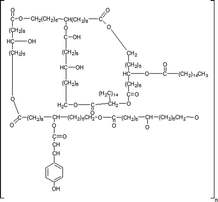
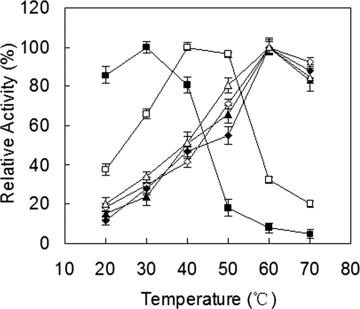
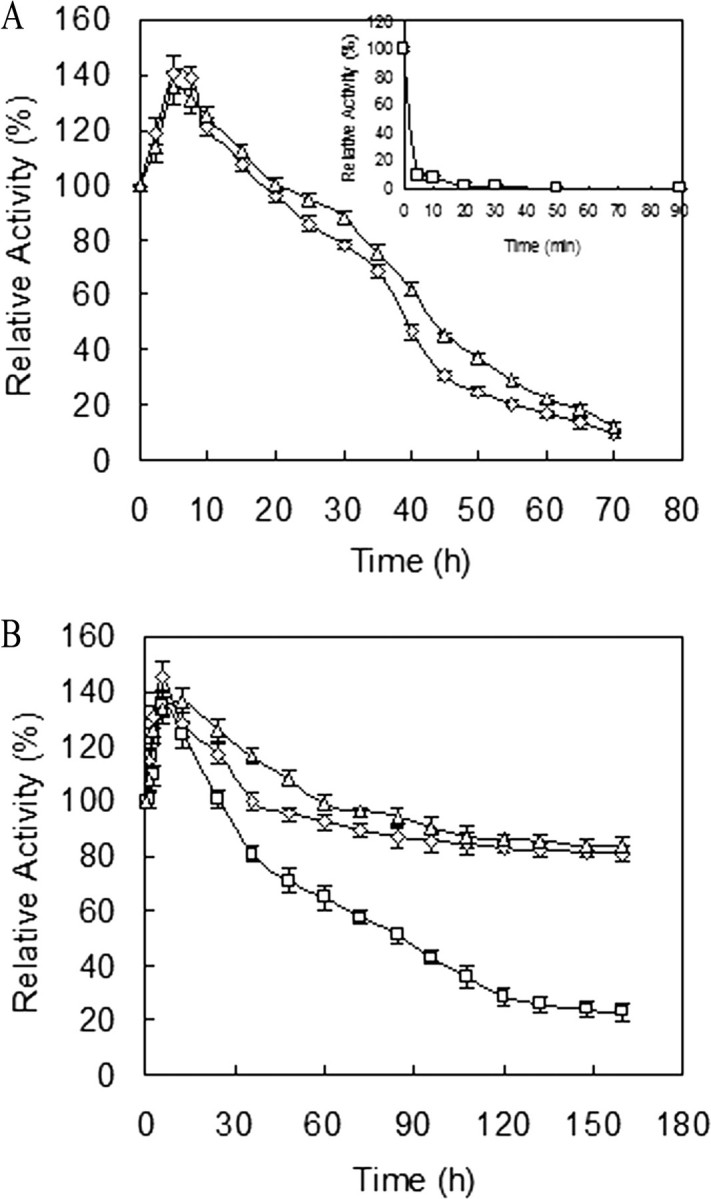
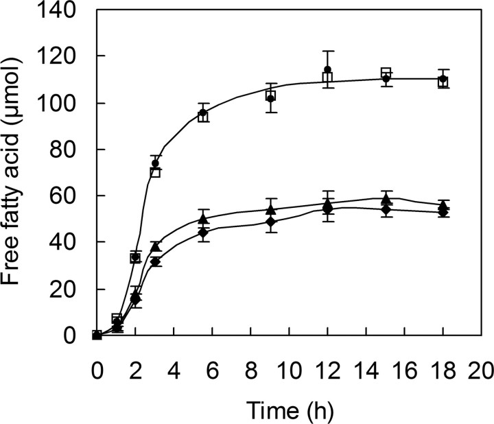
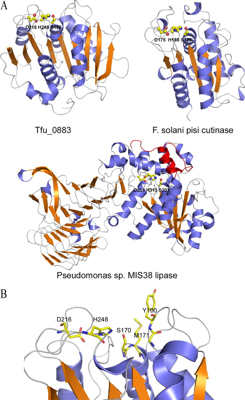
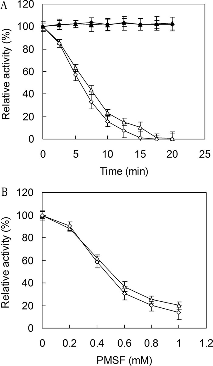
Cutinase, which exists in both fungi and bacteria, catalyzes the cleavage of the ester bonds of cutin. Fungal cutinases have been extensively studied, however, reports on bacterial cutinases have been limited due to the lack of knowledge concerning the identity of their open reading frames. In the present study, the cutinase from Thermobifida fusca was induced by cutin and purified to homogeneity by following p-nitrophenyl butyrate hydrolyzing activity. Peptide mass fingerprinting analysis of the wild-type enzyme matched two proteins, Tfu_0883 and Tfu_0882, which are 93% identical in sequence. Both proteins were cloned and overexpressed in their mature form. Recombinant Tfu_0883 and Tfu_0882 display very similar enzymatic properties and were confirmed to be cutinases by their capability to hydrolyze the ester bonds of cutin. Comparative characterization of Fusarium solani pisi and T. fusca cutinases indicated that they have similar substrate specificity and catalytic properties except that the T. fusca enzymes are thermally more stable. Homology modeling revealed that T. fusca cutinases adopt an alpha/beta-hydrolase fold that exhibits both similarities and variations from the fungal cutinase structure. A serine hydrolase catalytic mechanism involving a Ser(170)-His(248)-Asp(216) (Tfu_0883 numbering) catalytic triad was supported by active site-directed inhibition studies and mutational analyses. This is the first report of cutinase encoding genes from bacterial sources.


Figures

FIGURE 1.
FIGURE 1.
Structure of cutin.
FIGURE 2.
FIGURE 2.
Temperature optimum of cutinase. ▵ and ▴, Tfu_0882 using pNPB and triolein as substrates, respectively; ⋄ and ♦, Tfu_0883 using pNPB and triolein as substrates, respectively; □ and ▪, F. solani pisi cutinase using pNPB and triolein as substrates, respectively. Error bars correspond to the standard deviation of three determinations.
FIGURE 3.
FIGURE 3.
pH optimum of cutinase. ▵ and ▴, Tfu_0882 using pNPB and triolein as substrates, respectively; ⋄ and ♦, Tfu_0883 using pNPB and triolein as substrates, respectively; □ and ▪, F. solani pisi cutinase using pNPB and triolein as substrates, respectively. Enzyme activity was measured at 40 °C for F. solani pisi cutinase and 60 °C for Tfu_0882 and Tfu_0883, in either 25 mm potassium phosphate buffers (pH 6.0–7.0) or 20 mm Tris-HCl buffers (pH 7.0–9.0). Error bars correspond to the standard deviation of three determinations.
FIGURE 4.
FIGURE 4.
Thermostability of cutinase. The enzyme was incubated in 20 mm Tris-HCl (pH 8.0) at 60 °C (A) or 40 °C (B). ▵, ⋄, and □ represent the relative activity of Tfu_0882, Tfu_0883, and F. solani pisi cutinase, respectively. At various intervals, aliquots of the samples were removed and assayed for residual activity at their optimal temperature and pH. Error bars correspond to the standard deviation of three determinations.
FIGURE 5.
FIGURE 5.
Evaluation of possible synergistic effects between Tfu_0882 and Tfu_0883 in cutin degradation. Tfu_0882 (0.08 μmol) and Tfu_0883 (0.08 μmol) were together or individually incubated with 1% (w/v) apple cutin in 25 mm potassium phosphate buffer (pH 8) at 60 °C. The released fatty acids were quantified by titration with 0.02 n NaOH. ♦, Tfu_0883 alone; ▴, Tfu_0882 alone; •, Tfu_0882 and Tfu_0883; □, hypothetical sum of Tfu_0883 alone (♦) plus Tfu_0882 alone (▴). Error bars correspond to the standard deviation of three determinations.
FIGURE 6.
FIGURE 6.
Homology modeling of T. fusca cutinase. A, ribbon diagram of a predicted Tfu_0883 model compared with the structures of a fungal cutinase and a true bacterial lipase (Pseudomonas sp. MIS38 lipase). The catalytic triad residues are shown as ball-and-sticks. The lid domain of Pseudomonas sp. MIS38 lipase is shown in red. The Tfu_0883 model was predicted by SWISS-MODEL, an automated comparative protein modeling server (27). The template was SeL (PDB code 1jfr), which shares 63% sequence identity with Tfu_0883. B, the active site residues of T. fusca cutinase. The catalytic triad is formed by Ser170, His248, and Asp216. The oxyanion hole is formed by the main chain amides of Met171 and Tyr100.
FIGURE 7.
FIGURE 7.
Inactivation of cutinase by PMSF. A, time course of cutinase inactivation by PMSF. The enzymes were incubated with 1 mm PMSF (▵, Tfu_0882; ⋄, Tfu_0883) or without PMSF (▴, Tfu_0882; ♦, Tfu_0883) at 37 °C for various times and assayed for residual activity. B, concentration dependence of cutinase inactivation by PMSF. The enzymes (▵, Tfu_0882; ⋄, Tfu_0883) were incubated with various concentrations of PMSF at 37 °C for 10 min and assayed for residual activity. Error bars correspond to the standard deviation of three determinations.

References

    1. Fett, W. F., Gerard, H. C., Moreau, R. A., Osman, S. F., and Jones, L. E. (1992) Appl. Environ. Microb. 58 2123–2130 - PMC - PubMed
    1. Walton, T. J., and Kolattukudy, P. E. (1972) Biochemistry 11 1885–1896 - PubMed
    1. Purdy, R. E., and Kolattukudy, P. E. (1973) Arch. Biochem. Biophys. 159 61–69 - PubMed
    1. Gerard, H. C., Osman, S. F., Fett, W. F., and Moreau, R. A. (1992) Phytochem. Analysis 3 139–144
    1. Ferreira, B. S., Calado, C. R., van Keulen, F., Fonseca, L. P., Cabral, J. M., and da Fonseca, M. M. (2003) Appl. Microbiol. Biotechnol. 61 69–76 - PubMed

Publication types

LinkOut - more resources