. 2008 Aug;49(8):3677-86.
doi: 10.1167/iovs.07-1071.

Synaptic pathology in retinoschisis knockout (Rs1-/y) mouse retina and modification by rAAV-Rs1 gene delivery

Affiliations

Synaptic pathology in retinoschisis knockout (Rs1-/y) mouse retina and modification by rAAV-Rs1 gene delivery

Yuichiro Takada et al. Invest Ophthalmol Vis Sci. 2008 Aug.

Abstract

Purpose: At an early age, the retinoschisin knockout (Rs1-KO) mouse retina has progressive photoreceptor degeneration with severe disruption of the outer plexiform layer (OPL) that decreases at older ages. The electroretinogram (ERG) undergoes parallel changes. The b-wave amplitude from bipolar cells is reduced disproportionately to the photoreceptor a-wave at young but not at older ages. The protein expression and morphology of the OPL in Rs1-KO mice was investigated at different ages, to explore the role of the synaptic layer in these ERG changes.

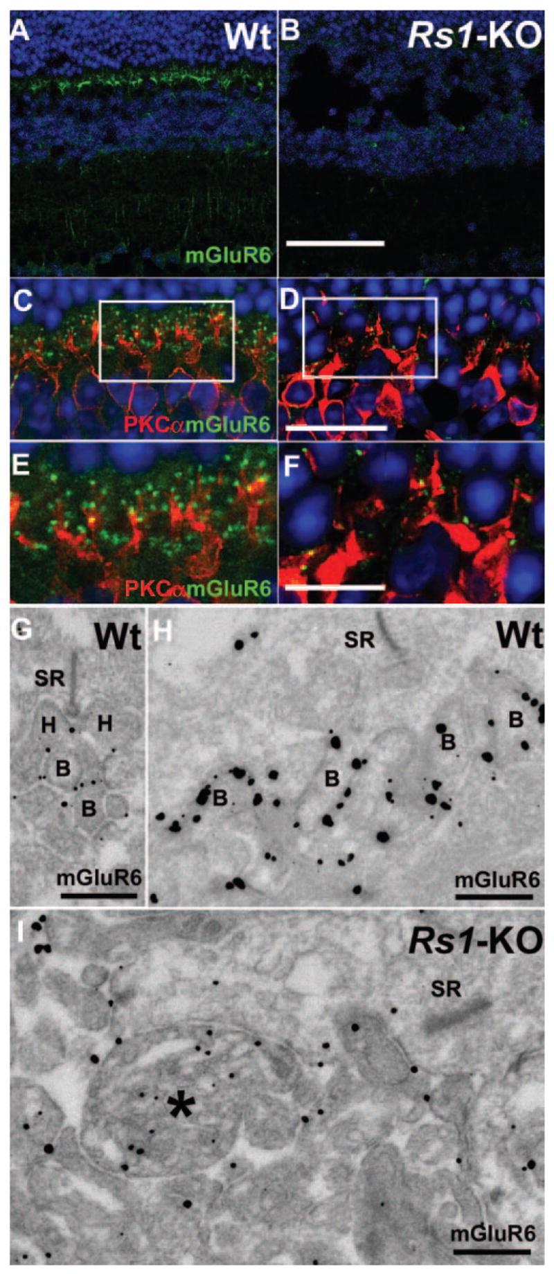
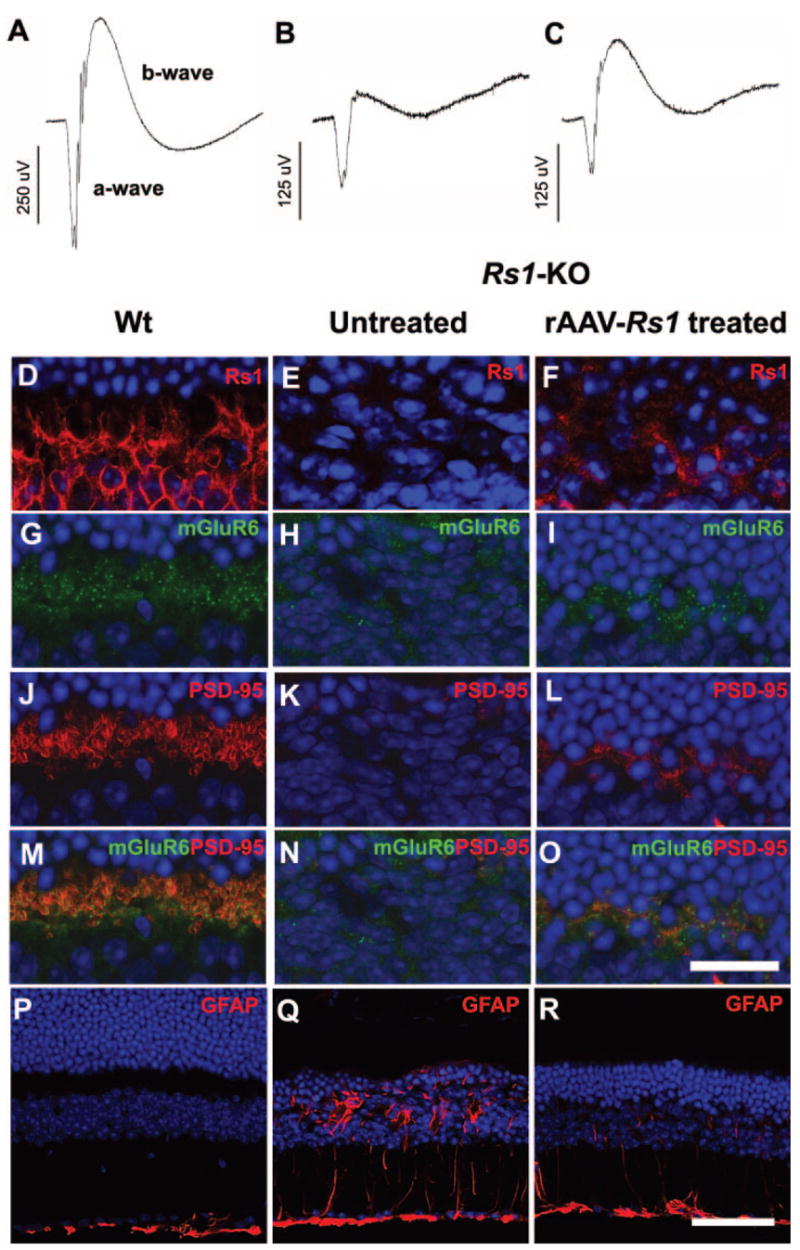
Methods: Retinas of wild-type (Wt) and Rs1-KO mice from postnatal day (P)7 to 12 months were evaluated by light and electron microscopy (EM) and biochemistry. PSD95 (postsynaptic density protein), mGluR6 (metabotropic glutamate receptor subtype 6), retinoschisin (Rs1), the Müller cell proteins glial fibrillary acidic protein (GFAP) and glutamine synthetase (GS), the bipolar cell marker protein kinase C alpha (PKCalpha), and the horizontal cell marker calbindin were localized by immunofluorescence and immuno-EM. Levels of PSD95 and mGluR6 were determined by quantitative Western blot. Rs1-KO mice treated by intravitreous injection of rAAV(2/2)-CMV-Rs1 in one eye at P14 were evaluated at 8 months by full-field scotopic ERG responses and retinal immunohistochemistry.

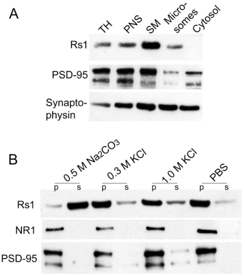
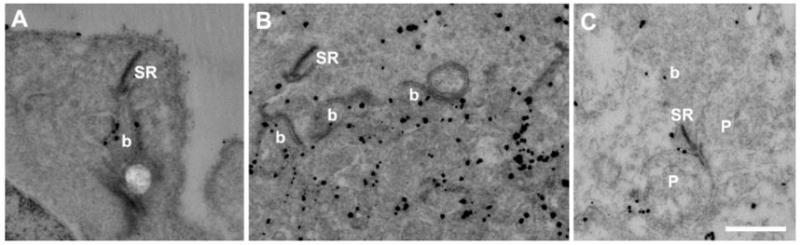
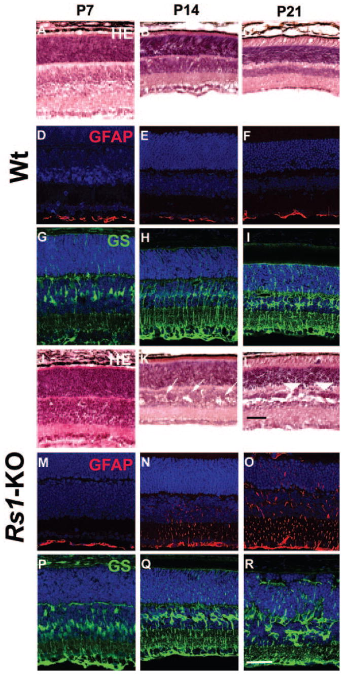
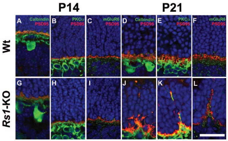
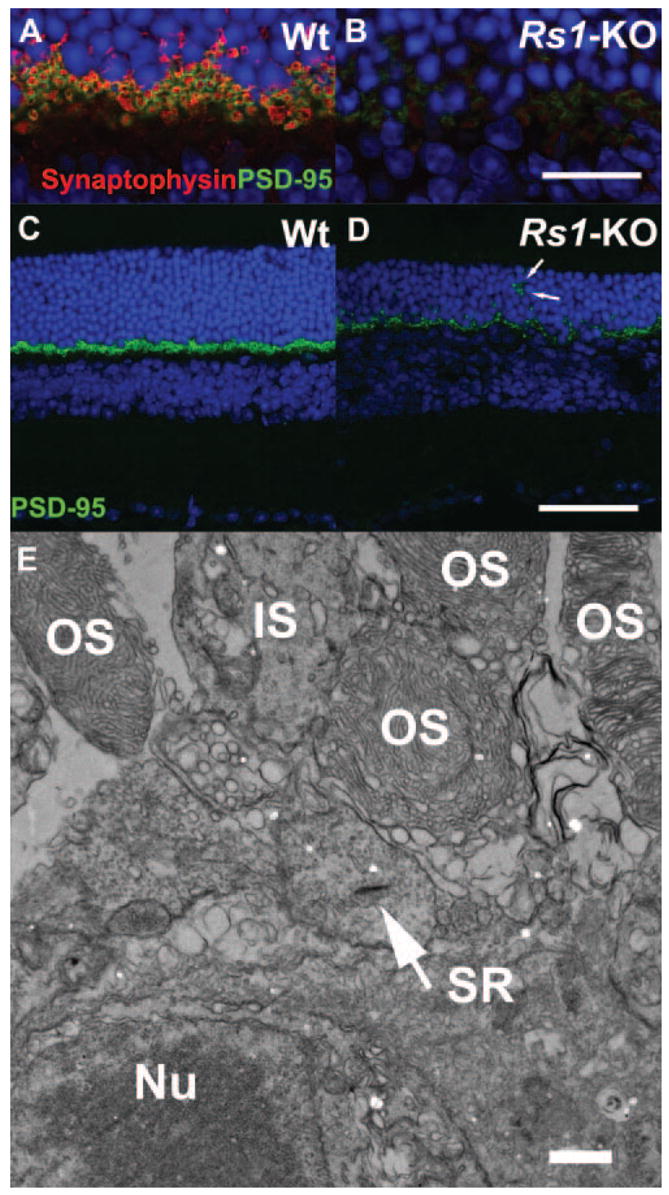
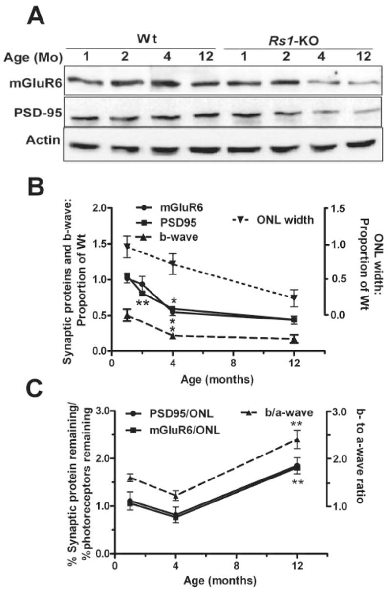
Results: Rs1 was associated with the outer surface of synaptic membranes in wild-type (Wt) retinas. PSD95 and mGluR6 were juxtaposed in the OPL of the Rs1-KO retinas by P14, implying that synaptic structures are formed. Light microscopic retinal morphology was similar in Wt and Rs1-KO at P14, but by P21, the OPL was disrupted in Rs1-KO, and some PSD95 and mGluR6 was mislocalized in the outer nuclear layer (ONL). GFAP expression spanned all retinal layers. EM showed synaptic structures adjacent to photoreceptor nuclei. PSD95 and mGluR6 levels were normal at 1 month on Western blot but declined to 59% (P < 0.001) and 55% (P < 0.05) of Wt, respectively, by 4 months. Levels thereafter showed no further reduction out to 12 months. Eyes injected with AAV-Rs1 were studied at 8 months by immunohistochemistry and had higher expression of PSD95 and mGluR6 and less GFAP expression compared with fellow untreated eyes.

Conclusions: In the Rs1-KO mouse, retinal layer formation and synaptic protein expression in the OPL is normal up to P14, implying normal development of synaptic connections. Aberrant localization of synaptic proteins by P21 indicates that displacement of developing and/or mature synapses contributes to the b-wave reduction at young ages, when photoreceptor numbers and synaptic protein levels are normal. The subsequent decline in PSD95 and mGluR6 between 1 and 12 months in Rs1-KO retina mirrors the course of b-wave change and provides evidence of causal relationship between the ERG and OPL changes. These findings and the improved structural integrity of the OPL and b-wave amplitude after Rs1 gene transfer therapy provide a cellular and molecular basis for interpreting the changes in retinal signaling in this model.


Figures

Figure 1
Figure 1
Biochemical localization of Rs1 to peripheral synaptic membranes. (A) SDS-PAGE of subcellular fractions from mouse retina followed by immunoblot analysis. The apparent molecular weight of the proteins: Rs1, monomer 26 kDa; PSD95, ~90 kDa; and synaptophysin, 38 kDa. TH, total homogenate; PNS, postnuclear supernatant; SM, synaptic membrane. (B) Alkaline and high-salt extraction of the synaptic membrane fraction. Synaptic membrane (30 μg protein) fractions were incubated in the presence of Na2CO3 or KCl or buffer alone (PBS). After high-speed centrifugation, the membrane-associated (pellets) and solubilized (supernatants) forms of Rs1 were analyzed by immunoblot analysis. The results are representative of three independent experiments pooling two eyes from one animal for each experiment.
Figure 2
Figure 2
Ultrastructural localization of Rs1 in the OPL and IPL. Electron micrographs of 2-month-old Wt retina immunostained with Rs1 antibody. Immunogold Rs1 particles were found in the OPL in rod spherules (A), cone pedicles (B), and bipolar cells. In the IPL, immunogold Rs1 particles were seen at the triad of bipolar and amacrine/ganglion cell processes (C). b, bipolar cell process; p, amacrine/ganglion process; SR, synaptic ribbon. Scale bar, 500 nm.
Figure 3
Figure 3
Retinal morphology and glial cell immunohistochemistry of Wt and Rs1-KO during postnatal development. Hematoxylin and eosin–stained histology of Wt (AC) and Rs1-KO (JL) retinas at P7, P14, and P21. At P7 (J) no abnormal features were observed in the Rs1-KO retina. By P14, several schisis cavities (arrows) were observed in the INL of Rs1-KO retinas (K). There were no major histologic abnormalities in the OPL. In the P21 Rs1-KO retina, the cavities were much more extensive in the INL and were also found in the OPL (arrowheads) (L). The vacuoles in the INL of Wt retinas at P7 are processing artifacts (A). Müller cells and GFAP immunohistochemical staining: Müller cells, stained with GS antibody were identified at developmental stages P7, P14, and P21 (Wt, GI; Rs1-KO, PR). In Wt retina, only the nerve fiber and ganglion cell layer showed GFAP labeling (DF). In Rs1-KO retina, GFAP expression at P7 was also restricted to the nerve fiber and ganglion cell layer, but between P14 (N) and P21 (O) GFAP labeling gradually extended across all layers, which is suggestive of Müller cell hypertrophy and reactive gliosis. Scale bar, 50 μm.
Figure 4
Figure 4
Presynaptic and postsynaptic terminal labeling in Wt and Rs1-KO retinas during postnatal development. PSD95 (red) labels the photoreceptor synaptic terminal; calbindin (green) labels horizontal cell; PKCα (green) labels rod bipolar cell processes; mGluR6 (green) labels bipolar cell terminals. In Wt retina at P14 and P21, terminals of horizontal (A, D) and rod bipolar cell processes (B, E) colocalized with PSD95 immunoreactivity in the OPL. Punctate labeling of mGluR6 (green) and presynaptic PSD95 (red) were juxtaposed to each other at P14 (C) and P21 (F) in Wt retinas. Rs1-KO retinas at P14 showed similar staining for synaptic markers PSD95/calbindin (G), PSD95/PKCα (H), and mGluR6 (I). However, at P21 the regular arrangement of PSD95 in the OPL was disrupted in Rs1-KO retinas and horizontal (J) and rod bipolar cell (K) processes extended into the ONL. Aberrant extension of mGluR6 into the ONL was seen at P21 in conjunction with mislocalized presynaptic photoreceptor axons (L). mGluR6 and PSD95 were juxtaposed to each other at these punctate ectopic locations in the ONL. Scale bar, 25 μm
Figure 5
Figure 5
Double labeling of photoreceptor synaptic terminals in 4-month-old Wt and Rs1-KO retinas. Wt and Rs1-KO mouse retinas from 4-month-old mice were stained with the presynaptic marker proteins synaptophysin (marking synaptic vesicles, red) and PSD95 (green). Labeling of both markers was greatly decreased in Rs1-KO retina (B, D) compared with Wt (A, C). In Rs1-KO, some PSD95 label was located in the ONL (D, arrows) indicating displaced terminals. At the ultrastructural level (E), electron microscopy confirmed displacement of photoreceptor terminals containing synaptic ribbons (SR, arrow) between the outer segments and nuclei of photoreceptor cells. OS, outer segment; IS, inner segment; Nu, Nuclei. Scale bar: (AD) 75 μm; (E) 100 nm.
Figure 6
Figure 6
Immunofluorescent staining of bipolar cell-specific markers in 4-month-old Rs1-KO retinas. Immunostaining intensity of bipolar cell mGluR6 (green) was greatly reduced in Rs1-KO mouse retina (B) compared with Wt at 4 months of age (A). Double labeling of Wt retina with PKCα (red) showed a regular network of rod bipolar cell processes colocalized with mGluR6 (C, E). In Rs1-KO mouse retina, PKCα labeling of bipolar cells dendrites are found in the ONL (D, F) along with sparse mGluR6 label (F). Ultrastructural localization of mGluR6 by immuno-EM showed it in the rod-bipolar (G) and cone-bipolar cell (H) plasma membrane of synaptic dendrites in 2-month-old Wt retina but mislocalized to the inside of bipolar cell dendrites (*) in Rs1-KO (I). Scale bar: (A, B) 75 μm; (C, D) 25 μm; (E, F) 12.5 μm; (G, I) 100 nm.
Figure 7
Figure 7
Immunoblot analysis of aging changes in mGluR6 and PSD95 protein expression in Wt and Rs1-KO mice retinas. Representative Western blots from Wt and Rs1-KO mice of 1, 2, 4, and 12 months of age. The targeted proteins were detected and quantified using enhanced chemiluminescence. At 1 and 2 months of age Rs1-KO synaptic protein levels appeared comparable to Wt. Both PSD95 and mGluR6 protein levels declined across 2 to 12 months. (B) Protein levels as a proportion of Wt at each age (solid lines). Intensity of mGluR6 and PSD95 bands was normalized to β-actin band intensity, and the Rs1-KO value was divided by the Wt value at each age. The points were obtained from three independent experiments using a pool of two to three retinas from different mice in each experiment. The effect of age and genotype was analyzed by two-way ANOVA and Bonferroni posttest. Differences in synaptic protein levels between Wt and Rs1-KO retina were statistically significant at 4 and 12 months (P < 0.01). Significant change from the previous age (top asterisk at each point, mGluR6; bottom asterisk at each point, PSD95: *P < 0.05, **P < 0.01, one-way ANOVA, Bonferroni posttest). For comparison to previously reported retinal morphology and ERG studies, the ONL cell count and b-wave amplitude, normalized by Wt at each age, are plotted on the same graph (dashed lines). (C) Comparison of postsynaptic response (b-wave) to synaptic protein changes with age normalized by photoreceptor responses (a-wave) or number (ONL count), respectively. The ERG values are from a previous study. **Significant change from the previous age (P < 0.01).
Figure 8
Figure 8
ERG retinal function and synaptic structure after Rs1 gene delivery to Rs1-KO retina. Representative dark-adapted ERG waveforms from an 8-month-old Rs1-KO mouse after gene delivery (B, C) show a larger b-wave in the treated eye, although it remains smaller than in Wt (A). Stimulus intensity = 0.6 log cd-s/m2. Expression of Rs1 (red) was seen in the OPL of Wt retina (D) and rAAV-Rs1 treated eye of the Rs1-KO mouse (F) but not in the untreated eye (E). Gene delivery elevated the expression levels of mGluR6 (green) and PSD95 (red) in OPL of the Rs1-KO retina (I, L, O) compared with the untreated fellow eye (H, K, N). Labeling of Wt retina for mGluR6, PSD95, and both mGluR6 and PSD95 is shown in (G), (J), and (M), respectively. Expression of GFAP (red) (P), was restricted to the inner limiting membrane in Wt but spanned the retina of Rs1-KO (Q). rAAV-mediated Rs1 gene delivery retina showed reduced GFAP protein expression (R). Scale bar: (DO) 25 μm; (PR) 75 μm.

References

    1. Sauer CG, Gehrig A, Warneke-Wittstock R, et al. Positional cloning of the gene associated with X-linked juvenile retinoschisis. Nat Genet. 1997;17:164–170. - PubMed
    1. Takada Y, Fariss RN, Tanikawa A, et al. A retinal neuronal developmental wave of retinoschisin expression begins in ganglion cells during layer formation. Invest Ophthalmol Vis Sci. 2004;45:3302–3312. - PubMed
    1. Takada Y, Fariss RN, Müller M, Bush RA, Rushing EJ, Sieving PA. Retinoschisin expression and localization in rodent and human pineal and consequences of mouse RS1 gene knockout. Mol Vis. 2006;12:1108–1116. - PubMed
    1. Vogel W. Discoidin domain receptors: structural relations and functional implications. FASEB J. 1999;13(suppl):S77–S82. - PubMed
    1. Wu WW, Wong JP, Kast J, Molday RS. RS1, a discoidin domain-containing retinal cell adhesion protein associated with X-linked retinoschisis, exists as a novel disulfide-linked octamer. J Biol Chem. 2005;280:10721–10730. - PubMed

Publication types

MeSH terms