. 2008 Jul;20(7):1805-17.
doi: 10.1105/tpc.108.059139. Epub 2008 Jul 25.

The EPIP peptide of INFLORESCENCE DEFICIENT IN ABSCISSION is sufficient to induce abscission in arabidopsis through the receptor-like kinases HAESA and HAESA-LIKE2

Affiliations

The EPIP peptide of INFLORESCENCE DEFICIENT IN ABSCISSION is sufficient to induce abscission in arabidopsis through the receptor-like kinases HAESA and HAESA-LIKE2

Grethe-Elisabeth Stenvik et al. Plant Cell. 2008 Jul.

Abstract

In Arabidopsis thaliana, the final step of floral organ abscission is regulated by INFLORESCENCE DEFICIENT IN ABSCISSION (IDA): ida mutants fail to abscise floral organs, and plants overexpressing IDA display earlier abscission. We show that five IDA-LIKE (IDL) genes are expressed in different tissues, but plants overexpressing these genes have phenotypes similar to IDA-overexpressing plants, suggesting functional redundancy. IDA/IDL proteins have N-terminal signal peptides and a C-terminal conserved motif (extended PIP [EPIP]) at the C terminus (EPIP-C). IDA can, similar to CLAVATA3, be processed by an activity from cauliflower meristems. The EPIP-C of IDA and IDL1 replaced IDA function in vivo, when the signal peptide was present. In addition, synthetic IDA and IDL1 EPIP peptides rescued ida and induced early floral abscission in wild-type flowers. The EPIP-C of the other IDL proteins could partially substitute for IDA function. Similarly to ida, a double mutant between the receptor-like kinases (RLKs) HAESA (HAE) and HAESA-LIKE2 (HSL2) displays nonabscising flowers. Neither overexpression of IDA nor synthetic EPIP or EPIP-C peptides could rescue the hae hsl2 abscission deficiency. We propose that IDA and the IDL proteins constitute a family of putative ligands that act through RLKs to regulate different events during plant development.


Figures

Figure 1.
Figure 1.
GUS Expression under the Control of the IDL Promoters. (A) IDL1:GUS expression in the columella root cap and in cells that are shed from the root. (B) to (D) Expression at the base of the pedicel, in the floral organ AZ, and in the funicle AZ, here represented by IDL2:GUS. (E) IDL3:GUS expression in vascular tissue of a young seedling. Expression in vascular tissue was also observed for IDL2:GUS, IDL4:GUS, and IDL5:GUS. (F) IDL4:GUS expression in guard cells. (G) GUS activity was seen in hydathodes both for IDL4:GUS and IDL5:GUS, here shown for IDL5:GUS.
Figure 2.
Figure 2.
Phenotype of Plants Overexpressing IDL Genes. (A) and (E) Wild-type Columbia (Col) floral organ AZs at positions 7 and 20 on the inflorescence. (B) and (F) 35S:IDA AZs at positions 7 and 20 showing premature abscission of floral organs and enlarged rounded AZ cells. (C) and (G) 35S:IDL1 AZs at positions 7 and 20. (D) and (H) 35S:IDL5 AZs at positions 7 and 20.
Figure 3.
Figure 3.
hae hsl2 Is Epistatic to 35S:IDA. (A) Position 12 siliques from wild-type Col, the hae hsl2 double mutant, the ida mutant, the 35S:IDA single locus homozygous line, and a hae hsl2 35S:IDA mutant plant. (B) RT-PCR performed on cDNA from AZ tissue of wild-type and hae hsl2 flowers (positions 4 to 8) using primers spanning the single intron of both HAE and HSL2, generating products of 120 and 1214 bp, respectively, from wild-type AZ cDNA, but not from hae hsl2 AZ cDNA. Genomic DNA controls (G) for HAE and HSL2 primers gave bands of 192 and 1301 bp, respectively. ACTIN2-7, giving a fragment of 255 bp with primers on each side of intron 2, was used as positive control. (C) pBS (i.e., the force required to remove petals from the flower) measured from positions 2 to 20 along the inflorescence of 15 wild-type (Col and C24), ida, and hae hsl2 plants. Standard deviations are shown as thin lines at the top of the columns. (D) hae hsl2 35S:IDA plant. Notice the abscission-deficient hae hsl2 phenotype. (E) HAE HSL2 35S:IDA plant with the 35S:IDA phenotype. Notice the early abscission and short siliques. (F) RT-PCR performed on cDNA from rosette leaf tissue of wild-type, hae hsl2, 35S:IDA, hae hsl2 35S:IDA, and a hae hsl2 plant without 35S:IDA from a segregating F2 population of a hae hsl2 35S:IDA cross. IDA primers amplified a fragment of 237 bp. ACTIN2-7 was used as positive control.
Figure 4.
Figure 4.
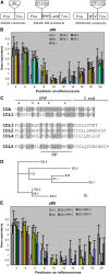
Constructs and pBS of Different IDA/IDL Recombinant Genes under the Control of IDA cis-Regulatory Elements. (A) Schematic presentation of various IDA:IDL constructs with IDA upstream (PIDA) and downstream (TIDA) cis-elements driving the expression. In the IDA:IDL gene swap constructs, the coding sequence of IDA has been exchanged with the coding sequence of IDL genes. In the IDA:IDL-IDA domain swap constructs, the IDA sequence encoding the signal peptide (SP) and variable region (V) has been exchanged with the corresponding IDL sequences. In the IDA:IDA-IDL domain swap constructs, the sequence encoding the EPIP-C of IDA has been exchanged with the corresponding IDL sequences. (B) pBS measured from positions 2 to 20 along the primary inflorescence of wild-type C24, the ida mutant, and ida mutant plants transformed with the gene swap constructs (cf. [A]) and for which full rescue of the ida mutant phenotype was not seen. Note reduced pBS compared with ida (i.e., partial rescue) at positions 12, 14, and 16. (C) Alignment of the C terminus of IDA and the five Arabidopsis IDLs. IDL1 is most similar to IDA and best-functioning. The middle cluster is the IDLs that can partially substitute for IDA, and at the bottom, IDL5, which cannot substitute for IDA, is shown. Amino acids in the EPIP-Cs of IDL identical to the IDA sequence are shaded; an asterisk indicates the EPIP residues common to IDA and IDL1 but not all the other IDLs. (D) Relationship between IDA and the IDL proteins. The phylogenetic tree was constructed using maximum likelihood analysis after alignment of the full-length protein sequences (see Supplemental Data Set 1 and Supplemental Figure 7 online). Figures indicate bootstrap values in percentages. (E) pBS for wild-type C24, the ida mutant, and the IDA:IDA-IDL EPIP-C swap constructs (cf. [A]) for which full rescue of the ida mutant phenotype was not seen. Note partial rescue (reduced pBS compared with ida) for several constructs at positions 8 to 20.
Figure 5.
Figure 5.
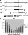
Deletion Analysis of IDA. (A) Schematic presentation of the IDA gene and three deletion constructs (IDAΔEPIP-C, where the sequence encoding EPIP-C had been deleted; IDAΔC-end, lacking the sequence encoding the C-end; and IDAΔVAR, where the sequence encoding the variable region had been removed). All the constructs contained the IDA upstream and downstream regulatory sequences (line with breaks) (Butenko et al., 2003). (B) pBS measured from positions 2 to 20 along the primary inflorescence for wild-type C24, ida, IDAΔVAR, and IDAΔEPIP-C.
Figure 6.
Figure 6.
Effects of Synthetic EPIP-C and EPIP Peptides on Abscission. (A) Percentage of abscised wild-type flowers (n = 30) after exposure to 10 μM EPIP IDA peptide (black) compared with control flowers (n = 30) without peptide in the medium (control, in green). (B) Percentage of abscised ida flowers (n = 30) after exposure to 10 μM EPIP IDA peptide (black) compared with ida flowers (n = 30) without peptide in the medium (control, in green). For (A) and (B), standard deviations based on three independent experiments are shown. (C) Floral AZ and pedicel AZ of wild-type flowers exposed to EPIP-C IDA or EPIP IDL1. Note secretion of white substance on floral AZ cells exposed to peptides and rupturing of the epidermal layer on pedicel AZ (arrow). The control had no peptide in the medium.
Figure 7.
Figure 7.
Proteolytic Processing of GST-Tagged IDAΔSP in Cauliflower Extracts. (A) GST-tagged IDA ΔSP or mCLV3 were untreated, incubated for 2 h in buffer, or incubated for 2 h with cauliflower (cfl) extracts. (B) Processing of GST-tagged IDAΔSP in cauliflower extracts in the presence of excessive amounts of His-tagged mCLV3 or BSA. Processed bands are indicated by an asterisk. Proteins were separated by SDS-PAGE and detected with anti-GST antibodies. (C) Alignment of IDA EPIP with MCLV3 and its preceding amino acid residues. Identical residues, conserved substitutions (asterisk), and semiconserved substitutions (colon) are indicated.

References

    1. Aalen, R.B., Butenko, M.A., Stenvik, G.E., Tandstad, N.M., and Patterson, S.E. (2006). Genetic control of floral abscission. In Floriculture, Ornamental and Plant Biotechnology: Advances and Topical Issues J.T. de Silva, ed (London: Global Science Books), pp. 101–108.
    1. Alonso, J.M., et al. (2003). Genome-wide insertional mutagenesis of Arabidopsis thaliana. Science 301 653–657. - PubMed
    1. Bleecker, A., and Patterson, S.E. (1997). Last exit: Senescence, abscission, and meristem arrest in Arabidopsis. Plant Cell 9 1169–1179. - PMC - PubMed
    1. Butenko, M.A., Patterson, S.E., Grini, P.E., Stenvik, G.E., Amundsen, S.S., Mandal, A., and Aalen, R.B. (2003). INFLORESCENCE DEFICIENT IN ABSCISSION controls floral organ abscission in Arabidopsis and identifies a novel family of putative ligands in plants. Plant Cell 15 2296–2307. - PMC - PubMed
    1. Butenko, M.A., Stenvik, G.E., Alm, V., Sæther, B., Patterson, S.E., and Aalen, R.B. (2006). Ethylene-dependent and -independent pathways controlling floral abscission are revealed to converge using promoter:reporter gene constructs in the ida abscission mutant. J. Exp. Bot. 57 3627–3637. - PubMed

Publication types

MeSH terms

Substances