. 2008 Nov;106(1):83-92.
doi: 10.1093/toxsci/kfn149. Epub 2008 Jul 27.

Abnormal liver development and resistance to 2,3,7,8-tetrachlorodibenzo-p-dioxin toxicity in mice carrying a mutation in the DNA-binding domain of the aryl hydrocarbon receptor

Affiliations

Abnormal liver development and resistance to 2,3,7,8-tetrachlorodibenzo-p-dioxin toxicity in mice carrying a mutation in the DNA-binding domain of the aryl hydrocarbon receptor

Maureen K Bunger et al. Toxicol Sci. 2008 Nov.

Abstract

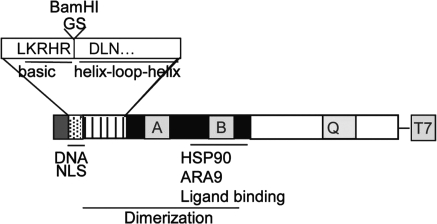
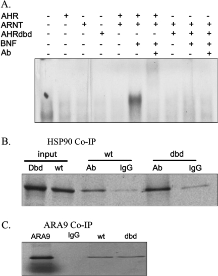
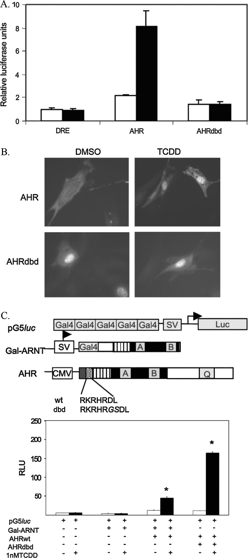
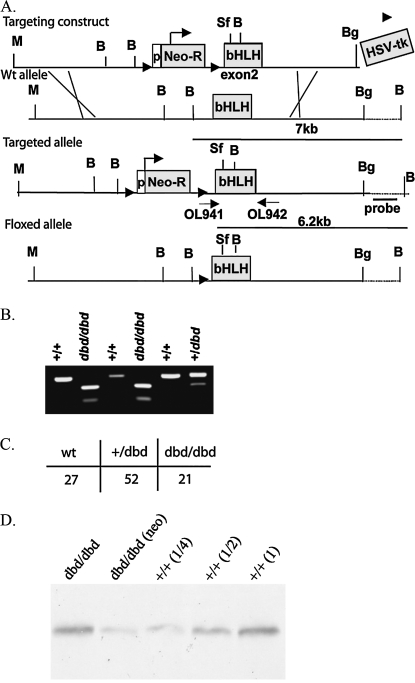
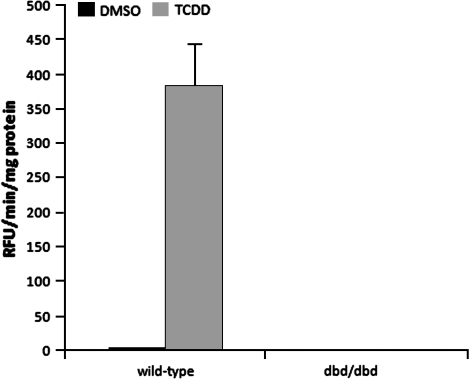
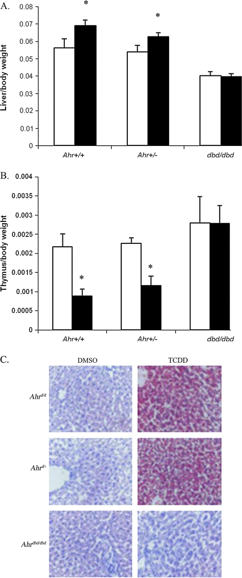
The aryl hydrocarbon receptor (AHR) is known for its role in the adaptive and toxic responses to a large number of environmental contaminants, as well as its role in hepatovascular development. The classical AHR pathway involves ligand binding, nuclear translocation, heterodimerization with the AHR nuclear translocator (ARNT), and binding of the heterodimer to dioxin response elements (DREs), thereby modulating the transcription of an array of genes. The AHR has also been implicated in signaling events independent of nuclear localization and DNA binding, and it has been suggested that such pathways may play important roles in the toxicity of 2,3,7,8-tetrachlorodibenzo-p-dioxin (TCDD). Here, we report the generation of a mouse model that expresses an AHR protein capable of ligand binding, interactions with chaperone proteins, functional heterodimerization with ARNT, and nuclear translocation, but is unable to bind DREs. Using this model, we provide evidence that DNA binding is required AHR-mediated liver development, as Ahr(dbd/dbd) mice exhibit a patent ductus venosus, similar to what is seen in Ahr(-/-) mice. Furthermore, Ahr(dbd/dbd) mice are resistant to TCDD-induced toxicity for all endpoints tested. These data suggest that DNA binding is necessary for AHR-mediated developmental and toxic signaling.


Figures

FIG. 1.
FIG. 1.
Schematic of the functional domains of the AHR protein. The enlarged portion depicts the GS insertion between the last residue of the basic domain and the first residue of the HLH domain, thereby introducing a BamHI restriction site.
FIG. 2.
FIG. 2.
Biochemical analysis of AHRdbd recombinant protein. (A) electromobility shift assay analysis of AHRdbd. AHR, ARNT, and AHRdbd proteins were expressed in reticulocyte lysate and equal quantities were incubated with a 32P-labeled, double-stranded oligo containing a single DRE consensus sequence. Shift of the AHR/ARNT heterodimer was induced by coincubation with 10μM BNF. An AHR-Ab was included to block the complex formation, controlling for specificity. (B,C) Co-IP of AHRdbd with HSP90. Wild-type or AHRdbd 35S-labeled in vitro–translated proteins were coincubated with reticulocyte lysate and HSP90-specific antibody (Ab) or preimmune IgG. Complexes were precipitated with Protein A-sepharose beads, separated on a 7.5% SDS-PAGE gel, and visualized with autoradiography. (D) Co-IP of AHRdbd with ARA9. T7-tagged AHR and AHRdbd were incubated with 35S-labeled ARA9. Complexes were precipitated using T7 antibody-coupled agarose beads and separated on a 7.5% SDS-PAGE gel.
FIG. 3.
FIG. 3.
Cellular characterization of the AHRdbd protein. (A) Luciferase assay for DRE-driven transcription. Ahr−/− 3T3 fibroblasts were transfected with equal amounts of DRE-Luc (PL256) and either AHR or AHRdbd recombinant cDNAs. Cells were then treated with 1nM TCDD (black bars) or 0.1% DMSO alone (white bars) for 24 h. Values represent relative luciferase units normalized to total protein levels. (B) Subcellular localization of AHRdbd. Indirect immunofluorescence was used to identify the subcellular localization of AHRdbd in Ahr−/− 3T3 fibroblasts transiently transfected with either Ahr+/+ or Ahrdbd/dbd. Prior to staining, nuclear translocation was induced by exposure of cells to 1nM TCDD for 2 h prior to staining. (C) Mammalian 2-hybrid analysis of AHRdbd interactions. The schematic diagram depicts the reporter construct (pG5luc), the “bait” construct (Gal-ARNT), and the “fish” construct (AHR), showing the amino acid sequence of the basic region in wild-type (wt) and AHRdbd (dbd) recombinant proteins. The two-hybrid analysis was carried out using equal amounts (0.33 μg) of transiently transfected Gal-ARNT and either wild-type AHR or AHRdbd, followed by incubation with 0.1% DMSO or 1nM TCDD. Values are expressed as relative luciferase units (*p < 0.001).
FIG. 4.
FIG. 4.
Generation of Ahrdbd mice. (A) Schematic diagram depicting the targeting construct used to generate the Ahrdbd allele in mice. Restriction enzyme sites shown are Mlu (M), BglII (Bg), BamHI (B), and SrfI (S). Arrowheads flanking the neomycin resistance cassette (Neo-R) indicate the location of LoxP sites. Shown in gray boxes are the locations of the Neo-R cassette, including the phosphoglycerate kinase promoter (p), the bHLH domain, and the Herpes Simplex Virus thymidine kinase gene cassette (HSV-tk). Primers used for PCR genotyping are shown (OL941 and OL942) as well as the location of the Southern probe used to genotype ES cells for recombination. The Floxed allele was generated by crossing the Ahrdbd Targeted allele animals to an animal expressing the Cre-recombinase protein driven by the CMV promoter, and the subsequent outcrossing to C57BL/6J to eliminate the Cre transgene. (B) PCR-based genotyping of Ahr+/+, dbd/dbd, and +/dbd mice. The amplified product from OL941 and OL942 is cut by BamHI only when the targeted allele is present. (C) Western blot analysis. AHR protein expression in liver extracts from wild-type and Ahrdbd/dbd mice. Lane 1: Floxed allele (Neo excised); lane 2: Targeted allele (Neo present); lane 3: Ahr+/+ (1/4 protein concentration); lane 4: Ahr+/+ (1/2 protein concentration); lane 5: Ahr+/+.
FIG. 5.
FIG. 5.
EROD analysis of liver microsomes from Ahrdbd/dbd mice. Wild-type 129SV/J and Ahdbd/dbd mice were administered a single injection of p-dioxane (-) or 32 μg/kg TCDD in p-dioxane (+) and sacrificed after 24 h. Microsomes were isolated from 0.5 g of liver, and EROD activity was quantified.
FIG. 6.
FIG. 6.
Developmental phenotype of Ahrdbd/dbd mice. (A) Relative organ wet weights of Ahrdbd/dbd mice (white bars) and wild-type littermates (black bars) sacrificed at 8 weeks of age (n = 5). *Indicates p < 0.01 by Student's t-test (wild-type versus Ahrdbd/dbd). (B) Representative H&E sections of livers from 7-day-old wild-type (littermate), Ahrdbd/dbd, and Ahr−/− mice (40× magnification). (C) Time-lapse angiography of wild-type (top row) and Ahrdbd/dbd (bottom row) littermates. Arrows identify key features as follows: BV, branching vessel; PV, portal vein; shIVC, suprahepatic inferior vena cava; ihIVC, infrahepatic inferior vena cava. Total time elapsed from the first panel to the last is approximately 10 s. (D) Incidence of patent DV in wild-type and Ahrdbd/dbd male mice as measured by trypan blue perfusion.
FIG. 7.
FIG. 7.
TCDD-induced phenotypic changes in Ahrdbd/dbd mice. (A) Hepatomegaly (expressed as relative liver weight) and (B) thymic involution (expressed as relative thymus weight) of DMSO- or TCDD-treated Ahr+/+ (n = 10), Ahr+/− (n = 11), and Ahrdbd/dbd (n = 10) mice as quantified 6 days after a single i.p. injection of p-dioxane (white bars) or 100 μg/kg TCDD (black bars). *Indicates p < 0.001. (C) Intrahepatic lipid accumulation. Frozen sections from DMSO- or TCDD-treated Ahr+/+, Ahr+/−, and Ahrdbd/dbd mice were stained with Oil Red-O (lipids, red) and hematoxylin (nuclei, blue).

References

    1. Bacsi SG, Hankinson O. Functional characterization of DNA-binding domains of the subunits of the heterodimeric aryl hydrocarbon receptor complex imputing novel and canonical basic helix-loop-helix protein-DNA interactions. J. Biol. Chem. 1996;271:8843–8850. - PubMed
    1. Berghard A, Gradin K, Pongratz I, Whitelaw M, Poellinger L. Cross-coupling of signal transduction pathways: The dioxin receptor mediates induction of cytochrome P-450IA1 expression via a protein kinase C-dependent mechanism. Mol. Cell. Biol. 1993;13:677–689. - PMC - PubMed
    1. Blankenship A, Matsumura F. 2,3,7,8-Tetrachlorodibenzo-p-dioxin-induced activation of a protein tyrosine kinase, pp60src, in murine hepatic cytosol using a cell-free system. Mol. Pharmacol. 1997;52:667–675. - PubMed
    1. Bunger MK, Moran SM, Glover E, Thomae TL, Lahvis GP, Lin BC, Bradfield CA. Resistance to 2,3,7,8-tetrachlorodibenzo-p-dioxin toxicity and abnormal liver development in mice carrying a mutation in the nuclear localization sequence of the aryl hydrocarbon receptor. J. Biol. Chem. 2003;278:17767–17774. - PubMed
    1. Buters JT, Doehmer J, Gonzalez FJ. Cytochrome P450-null mice. Drug Metab. Rev. 1999;31:437–447. - PubMed

Publication types

MeSH terms