. 2008 Aug;14(8):828-36.
doi: 10.1038/nm.1853. Epub 2008 Jul 27.

Hepatocyte-specific ablation of Foxa2 alters bile acid homeostasis and results in endoplasmic reticulum stress

Affiliations

Hepatocyte-specific ablation of Foxa2 alters bile acid homeostasis and results in endoplasmic reticulum stress

Irina M Bochkis et al. Nat Med. 2008 Aug.

Abstract

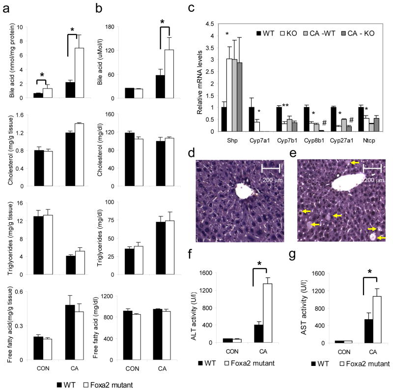
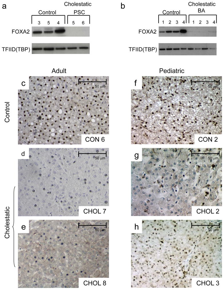
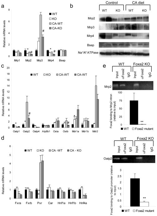
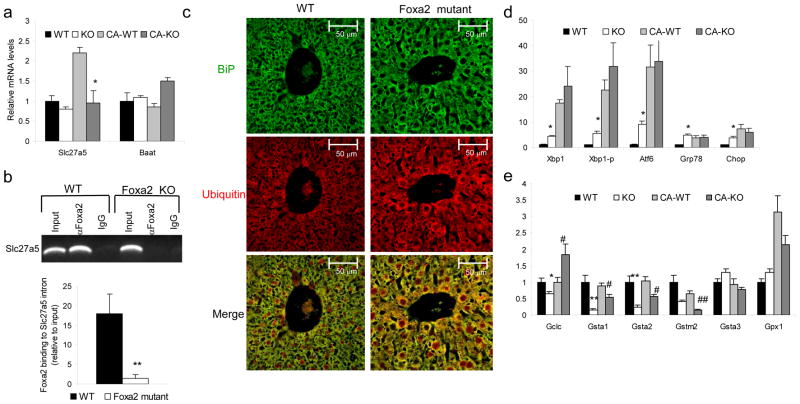
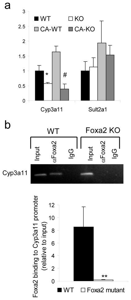
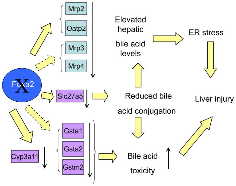
Production of bile by the liver is crucial for the absorption of lipophilic nutrients. Dysregulation of bile acid homeostasis can lead to cholestatic liver disease and endoplasmic reticulum (ER) stress. We show by global location analysis ('ChIP-on-chip') and cell type-specific gene ablation that the winged helix transcription factor Foxa2 is required for normal bile acid homeostasis. As suggested by the location analysis, deletion of Foxa2 in hepatocytes in mice using the Cre-lox system leads to decreased transcription of genes encoding bile acid transporters on both the basolateral and canalicular membranes, resulting in intrahepatic cholestasis. Foxa2-deficient mice are strikingly sensitive to a diet containing cholic acid, which results in toxic accumulation of hepatic bile salts, ER stress and liver injury. In addition, we show that expression of FOXA2 is markedly decreased in liver samples from individuals with different cholestatic syndromes, suggesting that reduced FOXA2 abundance could exacerbate the injury.


Conflict of interest statement

Conflict of interest: The authors have declared that no conflict of interest exists.

Figures

Figure 1
Figure 1
Foxa2 controls hepatic bile acid homeostasis and protects the liver from bile acid toxicity. (a) Hepatic bile acid levels are elevated two-fold in Foxa2 mutant mice (n = 6–7 animals per group) on regular chow, and increased an additional three-fold on bile acid diet relative to the wildtype control groups. (b) Serum bile acid concentrations are significantly increased in Foxa2loxP/loxPAlfp.Cre mice fed a bile acid diet compared with littermate controls (n = 4–5 animals per group). (c) Expression of Shp is elevated in Foxa2 mutant mice compared to the control littermates, while mRNA levels of Cyp7a1, Cyp7b1, Cyp8b1, Cyp27a1, and Ntcp are significantly reduced on standard diet (n = 7–8 animals per group). Expression of Shp increases equally for both groups on bile acid diet, while mRNA levels of Cyp7a1, Cyp7b1, Cyp8b1, Cyp27a1, and Ntcp decrease with cholic acid treatment. Representative liver sections from cholic acid-fed wildtype (d) and Foxa2loxP/loxPAlfp.Cre mice (e) stained with hematoxylin and eosin (H&E). Cholestatic injury is apparent on histological sections as indicated by increased hepatocyte dropout (arrows) in Foxa2-deficient livers. While serum alanine aminotransferase (ALT) (f) or aspartate aminotransferase (AST) (g) levels are not altered in Foxa2loxP/loxPAlfp.Cre mice on standard chow, the liver enzyme levels are disproportionately increased in the mutants on CA diet, significantly differing from those of their littermate controls. Values are represented as means plus standard error. P values were determined by Student’s t test. * p-value <0.05, WT vs. KO on standard diet, ** p-value <0.01, WT vs. KO on standard diet, # p-value<0.05 WT vs KO on CA diet.
Figure 2
Figure 2
FOXA2 expression is reduced in human cholestatic livers (a) Western blot analysis of protein nuclear extracts (30 μg) from three pediatric controls (corresponding to Control 3, 5, and 4 in Supplementary Table 3) and two patients with primary sclerosing cholangitis (corresponding to Cholestatic 5 and 6 in Supplementary Table 3) with antibodies against FOXA2 and TFIID (TBP) (loading control). FOXA2 protein levels were virtually undetectable in patients with primary sclerosing cholangitis. (b) Western blot analysis of protein nuclear extracts (30 μg) from four pediatric controls (corresponding to Control 1 through 4 in Supplementary Table 3) and four patients with biliary atresia (corresponding to Cholestatic 1 through 4) with antibodies against FOXA2 and TFIID (TBP) (loading control). FOXA2 protein levels were virtually undetectable in patients with biliary atresia. Immunohistochemical detection of FOXA2 protein in liver sections of adult (c) and pediatric (f) control and cholestatic patients (d–e, g–h). Labels in each panel correspond to patient numbers listed in Supplementary Table 3. While FOXA2 staining is clearly present in hepatocyte nuclei of the controls, FOXA2 presence is virtually absent in hepatocytes of the adult patients with primary sclerosing cholangitis (c, d) and pediatric patients with biliary atresia (g, h).
Figure 3
Figure 3
Foxa2 regulates several genes involved in hepatic bile acid transport directly. (a) Quantitative RT- PCR analysis for mRNA and (b) Western blot analysis of protein membrane fractions for expression of hepatic transporters. Both mRNA and protein levels of Mrp2 and Mrp4 are significantly decreased in Foxa2 mutants on both control and CA diet, while expression of Mrp3 is increased on standard chow but reduced on bile acid treatment in livers of Foxa2-deficient animals. (c) Expression of Oatp1 and Oatp2 is significantly reduced in Foxa2 mutants compared to controls on standard diet. mRNA levels of Oatp2 and Ostβ are decreased in livers of Foxa2-deficient mice on a diet supplemented with cholic acid. (d) Expression of numerous hepatic transcription factors involved in bile acid metabolism (n = 4 animals per group). While expression of Pxr is reduced 2-fold in Foxa2-deficient mice on standard diet, transcription of this gene is activated similarly on CA diet. mRNA levels of other DNA binding proteins do not differ between Foxa2 mutants and wildtype controls on either diet treatment. (e) Binding of Foxa2 to the Mrp2 intron and Oatp2 promoter is enriched relative to input chromatin in wildtype controls (n = 3 for each group). No promoter occupancy is seen in Foxa2-deficient liver, demonstrating the specificity of the assay for Foxa2. Values are represented as means plus standard error. P values were determined by Student’s t test. * p-value <0.05, **, p-value< 0.01, WT vs. KO on standard diet, # p-value<0.05, ## p-value <0.01, WT vs KO on CA diet
Figure 4
Figure 4
Foxa2 deficiency in the liver causes ER stress.. (a) Expression of Slc27a5 in liver RNA of wild-type and Foxa2loxP/loxPAlfp.Cre mice shows a 2-fold decrease in Foxa2 mutants as compared to their control littermates on cholic acid diet, while expression of Baat, or bile acid CoA amino acid N-acyltransferase, is not changed (n = 4 animals for each group). (b) ChIP analysis of promoter occupancy in liver chromatin from wild-type or Foxa2loxP/loxPAlfp.Cre mice (n = 3 for each group). The second exon amplicon of Slc27a5 is enriched 20-fold relative to input DNA in liver chromatin from wildtype mice. This binding is completely lost in the absence of Foxa2. (c) Immunofluorescence staining of liver sections from control and mutant mice using antibodies against BiP (top panel) and ubiquitin (middle panel). Mutant liver sections exhibit elevated levels of the chaperone protein BiP, as well as elevated cytosolic ubiquitin (middle panel), where aberrant ER proteins are translocated to be degraded by the ubiquitin-proteasome system. The merge of the two confocal images demonstrates increased co-localization of BiP and ubiquitin in livers of Foxa2loxP/loxP;Alfp.Cre mice (bottom panel). (d) Expression of ER stress markers in liver RNA of wild-type and Foxa2loxP/loxPAlfp.Cre mice (n = 4 animals per group). (e) Q-PCR analysis for messenger RNA of hepatic glutathione enzymes (n = 4 animals per group). Values are represented as means plus standard error. P values were determined by Student’s t test. * p-value <0.05, **, p-value< 0.01, WT vs. KO on standard diet, # p-value<0.05, ## p-value <0.01, WT vs KO on CA diet
Figure 5
Figure 5
Foxa2 regulates the phase I detoxification enzyme, Cyp3a11. (a) Quantitative RT-PCR analysis for expression of Cyp3a11 in liver RNA of wild-type and Foxa2loxP/loxPAlfp.Cre mice shows a reduction of 2-fold on standard diet and 5-fold decrease on cholic acid diet in the absence of Foxa2, while expression of Sult2a1, a phase II detoxification enzyme is not changed (n = 4 animals for each group). Values are represented as means plus standard error. P values were determined by Student’s t test. * p-value <0.05, WT vs. KO on standard diet, # p-value<0.05 WT vs. KO on CA diet (b) ChIP analysis of promoter occupancy evaluated by conventional and quantitative PCR in liver chromatin from wild-type or Foxa2loxP/loxPAlfp.Cre mice (n = 3 for each group). The Cyp3a11 promoter amplicon is enriched 8-fold relative to input DNA in liver chromatin from wildtype mice. This binding is completely lost in the absence of Foxa2. Values are represented as means plus standard error. P values were determined by Student’s t test. **, p -value< 0.01.
Figure 6
Figure 6
A model for the regulation of hepatic bile acid homeostasis by Foxa2. Foxa2 regulates the expression of Mrp2 and Oatp2 directly and that of Mrp3 and Mrp4 indirectly. Mrp3 and Mrp4 facilitate export of bile acids from hepatocytes into circulation, the organic anion carrier Oatp2 is involved in sodium-independent hepatic uptake of bile salts, while Mrp2 is involved in secretion of bile acids and their anionic conjugates into bile. Expression of conjugation enzymes Slc27a5, Gsta1, Gsta2, and Gstm2 also depends on Foxa2. Reduced conjugation of bile acids coupled with decreases in mRNA levels of the transporters due to absence of Foxa2 leads to elevated hepatic bile acids, which in turn cause ER stress. Expression of Cyp3a11 is directly controlled by Foxa2 as well. Cyp3a11 is important in hydroxylation of the hydrophobic bile acids, protecting the liver from bile acid toxicity. Together, ER stress and increased bile acid toxicity in Foxa2loxP/loxPAlfp.Cremice lead to liver injury.

References

    1. Kaestner KH. The hepatocyte nuclear factor 3 (HNF3 or FOXA) family in metabolism. Trends Endocrinol Metab. 2000;11:281–5. - PubMed
    1. Costa RH, Grayson DR, Darnell JE., Jr Multiple hepatocyte -enriched nuclear factors function in the regulation of transthyretin and alpha 1-antitrypsin genes. Mol Cell Biol. 1989;9:1415–25. - PMC - PubMed
    1. Ang SL, et al. The formation and maintenance of the definitive endoderm lineage in the mouse: involvement of HNF3/forkhead proteins. Development. 1993;119:1301–15. - PubMed
    1. Monaghan AP, Kaestner KH, Grau E, Schutz G. Postimplantation expression patterns indicate a role for the mouse forkhead/HNF-3 alpha, beta and gamma genes in determination of the definitive endoderm, chordamesoderm and neuroectoderm. Development. 1993;119:567–78. - PubMed
    1. Lee CS, Friedman JR, Fulmer JT, Kaestner KH. The initiation of liver development is dependent on Foxa transcription factors. Nature. 2005;435:944–7. - PubMed

Publication types