Comparative Study
. 2008 Sep;17(9):1230-6.
doi: 10.1007/s00586-008-0725-7. Epub 2008 Jul 26.

Preoperative imaging of cervical pedicles: comparison of accuracy of oblique radiographs versus axial CT scans

Affiliations
Comparative Study

Preoperative imaging of cervical pedicles: comparison of accuracy of oblique radiographs versus axial CT scans

James P Sieradzki et al. Eur Spine J. 2008 Sep.

Abstract

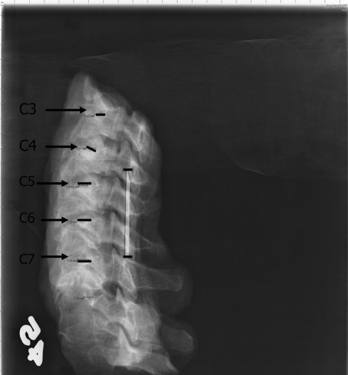
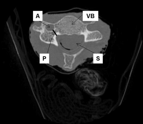
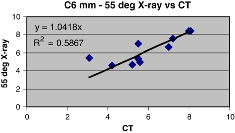
In spite of concerns about safety during their insertion, cervical spine pedicle screws have demonstrated biomechanical superiority over lateral mass screws in several biomechanical studies. One of the concerns for placement of cervical pedicle screws is their small size. Preoperative planning with computed tomography to assess pedicle width has been shown to be extremely accurate and is recommended by several authors. To date there has been no study assessing the accuracy of oblique radiographs for pedicle measurement. We sought to compare accuracy of the oblique radiographic measurements of cervical pedicle width with axial CT scan measurements. Five fresh-frozen human cadaveric cervical spines C3-C7 were studied. Thin cut 1.25 mm computed tomography axial cuts were made through the pedicle isthmus. Oblique radiographs at 35 degrees , 45 degrees , and 55 degrees angles were taken of the right and left pedicles of each specimen using a standardized technique. Each radiograph contained a pin of known length to correct for magnification. All pedicles were again measured and corrected for magnification using the standard pin. Corrected oblique radiograph measurements were compared to CT for each specimen. The outer pedicle width was measured and agreed upon by consensus. The radiograph measurements were on average significantly larger than CT measurements for the pedicles indicating that the pin standard did not completely correct magnification. Plain radiographic data failed to reveal that one oblique angle was favorable to another in terms of magnification or precision. Plain radiographs at oblique angles do not provide accurate measurements of subaxial cervical pedicles at 35 degrees , 45 degrees , or 55 degrees angles. We recommend that thin cut axial CT scans be obtained on all patients prior to transpedicular fixation in the cervical spine.


Figures

Fig. 1
Fig. 1
Experimental setup for oblique radiographs with saw bones cervical model in place, black arrow points to position of standard pin. Height of the board was altered using standard pegs in order to create 35°, 45°, and 55° angle from the table below
Fig. 2
Fig. 2
45° oblique radiograph demonstrating the pedicle measurements C3–C7 obtained with the PACS workstation, arrows point to digital measurements of each pedicle. Measurement of standard pin is evident as well
Fig. 3
Fig. 3
Axial cut of the CT scan where the widest portion of the pedicle isthmus was measured. The outer diameter of the pedicle was measured as shown. The vertebral artery (A), spinal canal (S), vertebral body (VB) and pedicle (P) are labeled
Fig. 4
Fig. 4
CT and 55° oblique radiograph measurement plot demonstrating lack of correlation with R2 = 0.59. Oblique measurements for each cervical level C3–C7 were plotted against CT values with similar results not shown here. None of the obliques at any cervical level correlated with CT measurements
Fig. 5
Fig. 5
Correction factors plotted against radiographic angle. Correction increases as radiographic angle according to experimental setup. Additionally the correction factors for the right pedicles were consistently greater consistent with pin placement
Fig. 6
Fig. 6
Percent magnification plotted against height of pin from radiographic plate demonstrating linear increase in magnification

References

    1. Abumi K, Ito M, Taneichi H, Kaneda K. Transpedicular screw fixation for traumatic lesions of the middle and lower cervical spine: description of the techniques and preliminary report. J Spinal Disord. 1994;7:19–28. doi: 10.1097/00002517-199407010-00003. - DOI - PubMed
    1. Abumi K, Kaneda K. Pedicle screw fixation for nontraumatic lesions of the cervical spine. Spine. 1997;22(16):1853–1863. doi: 10.1097/00007632-199708150-00010. - DOI - PubMed
    1. Abumi K, Shono Y, Taneichi H, Ito M, Kaneda K. Correction of cervical kyphosis using pedicle screw fixation systems. Spine. 1999;24:2389–2396. doi: 10.1097/00007632-199911150-00017. - DOI - PubMed
    1. Abumi K, Shono Y, Ito M, Taneichi H, Kotani Y, Kaneda K. Complications of pedicle screw fixation in reconstructive surgery of the cervical spine. Spine. 2000;25:962–969. doi: 10.1097/00007632-200004150-00011. - DOI - PubMed
    1. Abumi K, Takada T, Shono Y, Kaneda K, Fujiya M. Posterior occipitocervical reconstruction using cervical pedicle screws and platerod systems. Spine. 1999;24:1425–1434. doi: 10.1097/00007632-199907150-00007. - DOI - PubMed

Publication types