. 2009 May;30(5):1580-91.
doi: 10.1002/hbm.20625.

Decreased basal fMRI functional connectivity in epileptogenic networks and contralateral compensatory mechanisms

Affiliations

Decreased basal fMRI functional connectivity in epileptogenic networks and contralateral compensatory mechanisms

Gaelle Bettus et al. Hum Brain Mapp. 2009 May.

Abstract

A better understanding of interstructure relationship sustaining drug-resistant epileptogenic networks is crucial for surgical perspective and to better understand the consequences of epileptic processes on cognitive functions. We used resting-state fMRI to study basal functional connectivity within temporal lobes in medial temporal lobe epilepsy (MTLE) during interictal period. Two hundred consecutive single-shot GE-EPI acquisitions were acquired in 37 right-handed subjects (26 controls, eight patients presenting with left and three patients with right MTLE). For each hemisphere, normalized correlation coefficients were computed between pairs of time-course signals extracted from five regions involved in MTLE epileptogenic networks (Brodmann area 38, amygdala, entorhinal cortex (EC), anterior hippocampus (AntHip), and posterior hippocampus (PostHip)). In controls, an asymmetry was present with a global higher connectivity in the left temporal lobe. Relative to controls, the left MTLE group showed disruption of the left EC-AntHip link, and a trend of decreased connectivity of the left AntHip-PostHip link. In contrast, a trend of increased connectivity of the right AntHip-PostHip link was observed and was positively correlated to memory performance. At the individual level, seven out of the eight left MTLE patients showed decreased or disrupted functional connectivity. In this group, four patients with left TLE showed increased basal functional connectivity restricted to the right temporal lobe spared by seizures onset. A reverse pattern was observed at the individual level for patients with right TLE. This is the first demonstration of decreased basal functional connectivity within epileptogenic networks with concomitant contralateral increased connectivity possibly reflecting compensatory mechanisms.


Figures

Figure 1
Figure 1
Regions of Interest (ROIs) defined in bilateral temporal lobes used to extract fMRI signal time courses during resting periods. ROIs (posterior hippocampus: brown; anterior hippocampus: red; entorhinal cortex: orange; amygdala: yellow; temporal pole (Brodmann area 38): blue) were automatically defined on a digital Talairach atlas and applied on the coregistered and spatially normalized EPI images for each subject.
Figure 2
Figure 2
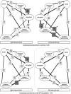
Basal functional connectivity of the medial temporal lobes in controls (top) and in left MTLE patients (bottom). Connecting lines correspond to significant basal functional connectivity. Links thickness reflects correlation values. Normalized correlation coefficients and standard deviation (brackets) are indicated in squares. In patients, epileptogenic zone is outlined. Hemispheric predominance of correlation coefficients are marked by grey background squares (uncorrected P < 0.05) and stars stand for surviving P values to multiple comparison correction (Mann Whitney test, corrected P < 0.005).
Figure 3
Figure 3
Differences in basal functional connectivity between the left MTLE patients and controls. Relative to controls, MTLE patients showed disruption (entorhinal cortex––anterior hippocampus, crossed dashed line) and decreased functional connectivity (anterior–posterior hippocampi, dotted line) inside the left temporal lobe and increased basal functional connectivity inside the right temporal lobe (anterior–posterior hippocampi, bold line) (Mann Whitney test, corrected P < 0.005). Epileptogenic zone is outlined.
Figure 4
Figure 4
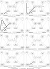
Individual patterns of basal functional connectivity observed in left MTLE patients. Links with normal basal functional connectivity values are displayed in grey. Relative to the mean and standard deviation of controls, patients have been classified as abnormal for z‐score values above or below 2 SD. Increased connectivity are reported in bold and decreased connectivity with crossed dashed line. One patient had a normal profile. Seven patients had at least one decreased or disrupted connection. Four patients had at least one increased connection in the right temporal lobe.
Figure 5
Figure 5
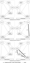
Individual patterns of basal functional connectivity observed in right MTLE patients. Links with normal basal functional connectivity values are displayed in grey. Relative to the mean and standard deviation of controls, patients have been classified as abnormal for z‐score values above or below 2 SD. Increased connectivity are reported in bold and decreased connectivity with crossed dashed line. Two patients had interrupted connectivity in one or both lobes. Two patients had at least one increased connection in the left lobe.
Figure 6
Figure 6
Reorganization of functional connectivity in left MTLE patients. Correlation between basal functional connectivity values between the right anterior and posterior hippocampi and the extent of disconnection inside the left hemisphere reflected by the number of links with abnormal connectivity values (Spearman Rho = 0.792, P = 0.019).
Figure 7
Figure 7
Compensatory effects of functional reorganization in left MTLE patients. Significant positive correlation (R = 0.711, P = 0.048) between the increase in basal functional connectivity values between the right anterior and posterior hippocampi link and the memory scores (working memory quotients of the WMS III battery).

References

    1. Addis DR,Moscovitch M,McAndrews MP ( 2007): Consequences of hippocampal damage across the autobiographical memory network in left temporal lobe epilepsy. Brain 130(Pt 9): 2327–2342. - PubMed
    1. Baas D,Aleman A,Kahn RS ( 2004): Lateralization of amygdala activation: A systematic review of functional neuroimaging studies. Brain Res Brain Res Rev 45: 96–103. - PubMed
    1. Bartels A,Zeki S ( 2005): The chronoarchitecture of the cerebral cortex. Philos Trans R Soc Lond B Biol Sci 360: 733–750. - PMC - PubMed
    1. Bartolomei F,Wendling F,Bellanger JJ,Regis J,Chauvel P ( 2001): Neural networks involving the medial temporal structures in temporal lobe epilepsy. Clin Neurophysiol 112: 1746–1760. - PubMed
    1. Bartolomei F,Wendling F,Regis J,Gavaret M,Guye M,Chauvel P ( 2004): Preictal synchronicity in limbic networks of mesial temporal lobe epilepsy. Epilepsy Res 61: 89–104. - PubMed

Publication types