. 2008 Oct;64(4):465-70.
doi: 10.1002/ana.21449.

Sustained improvement of spinal muscular atrophy mice treated with trichostatin A plus nutrition

Affiliations

Sustained improvement of spinal muscular atrophy mice treated with trichostatin A plus nutrition

Heather L Narver et al. Ann Neurol. 2008 Oct.

Abstract

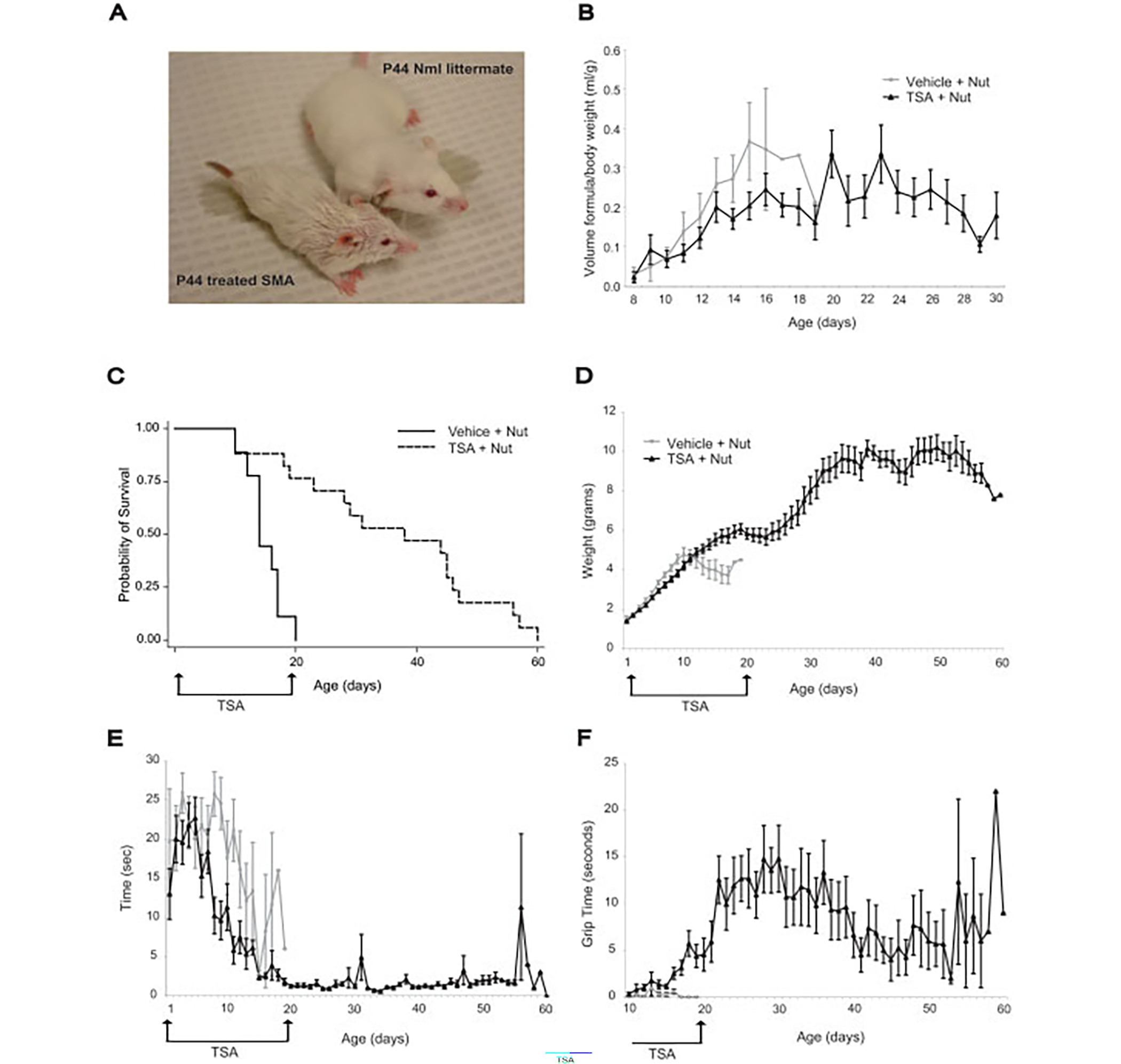
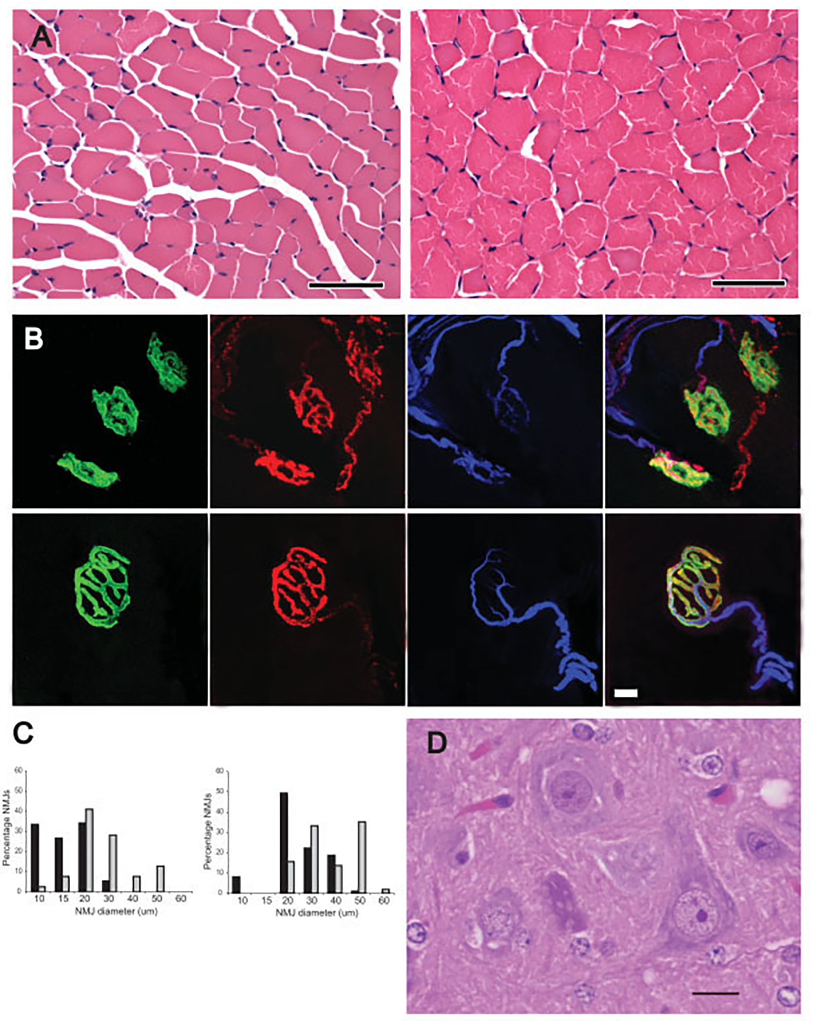
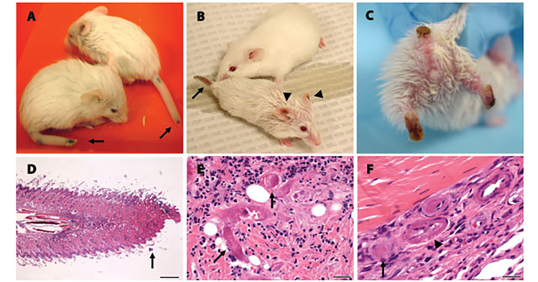
Early treatment with the histone deacetylase inhibitor, trichostatin A, plus nutritional support extended median survival of spinal muscular atrophy mice by 170%. Treated mice continued to gain weight, maintained stable motor function, and retained intact neuromuscular junctions long after trichostatin A was discontinued. In many cases, ultimate decline of mice appeared to result from vascular necrosis, raising the possibility that vascular dysfunction is part of the clinical spectrum of severe spinal muscular atrophy. Early spinal muscular atrophy disease detection and treatment initiation combined with aggressive ancillary care may be integral to the optimization of histone deacetylase inhibitor treatment in human patients.


Figures

Fig 1.
Fig 1.
Early trichostatin A (TSA) treatment combined with nutritional support substantially improves survival and results in sustained improvement in motor function. (A) TSA-treated spinal muscular atrophy (SMA) mouse and normal littermate at postnatal day 44 (P44). The SMA mouse is smaller than the normal littermate but is upright and ambulates well. The rough coat is secondary to antibiotic ointment. (B) SMA mice treated with vehicle (n = 9; gray lines) and TSA (n = 17; black lines) consumed approximately equal volumes of formula (ml/gram of body weight). TSA-treated SMA mice continued to receive formula feedings until the fifth week of life. (C) SMA mice treated with TSA (P2-P20) plus nutrition (dashed line) had a substantial increase in median survival of 170% (38 days) compared with mice treated with vehicle plus nutrition (14 days; solid line) (log rank < 0.001). (D) SMA mice receiving TSA plus nutrition (black lines) had weight gain long after TSA was discontinued. Gray lines represent vehicle plus nutrition. (E) SMA mice receiving TSA plus nutrition (black lines) showed improved righting time during the first 2 weeks and maintained this ability thereafter. Gray lines represent vehicle plus nutrition. This difference is significant at P8 (p < 0.001). (F) SMA mice receiving TSA plus nutrition (black lines) showed improved forelimb grip time. Gray lines represent vehicle plus nutrition. This difference is significant at P13 (p = 0.04).
Fig 2.
Fig 2.
Long-lived trichostatin A (TSA)–treated spinal muscular atrophy (SMA) mice have morphologically immature but connected motor units. (A) Myofibers of the TA muscle were hypotrophic, but otherwise morphologically normal in a treated postnatal day 44 (P44) SMA mouse (left) compared with an age-matched normal littermate (right). (B) Triple staining of neuromuscular junctions (NMJs) with α-bungarotoxin (green), synaptophysin (red), and neurofilament (blue). NMJs in the paraspinal muscle of a treated P46 SMA mouse (top) were small and simplified compared with a P46 normal littermate. (C) As indicated in the Table, there was minimal evidence of denervated NMJs in the TA, intercostal, or paraspinal muscles of P39 and P46 SMA mice compared with normal littermates (NL; gray bars). Only the SMA P39 (black bars) TA muscle showed a statistically significant increase in denervation (*p = 0.005). As indicated in histograms, NMJ diameter was reduced in the paraspinal muscles of treated SMA mice compared with age-matched normal littermates. (D) Many morphologically normal anterior horn cells were present in ventral horn of lumbar spine of a treated P29 SMA mouse. Scale bars 50μm (A); 10μm (B); 30μm (C).
Fig 3.
Fig 3.
Long-lived trichostatin A (TSA)–treated spinal muscular atrophy (SMA) mice show progressive distal necrosis and small-vessel thrombosis. (A) Two postnatal day 25 (P25) SMA mice showing early necrosis at the tips of the tails. (B) A P44 SMA mouse and age-matched normal littermate. The SMA mouse shows necrosis of the tail (arrow) and ears (arrowheads). (C) A P47 SMA mouse showing severe tail and bilateral hind-limb necrosis resulting in significant tissue loss. (D) Hematoxylin-and-eosin–stained longitudinal section of the tail from a P29 SMA mouse demonstrated necrosis at the tail tip. Scale bar = 400μm. (E) High-power view of the tail just proximal to the junction of viable and necrotic tissue. Arrows highlight two thrombosed small vessels. Scale bar = 25μm. (F) High-power view of foot from a P57 SMA mouse showing four vessels. One vessel indicated by the arrow is thrombosed and one vessel indicated by the arrowhead shows fibrinoid necrosis in the vessel wall.

References

    1. Lefebvre S, Burglen L, Reboullet S, et al. Identification and characterization of a spinal muscular atrophy-determining gene. Cell 1995;80:155–165. - PubMed
    1. Lorson CL, Hahnen E, Androphy EJ, Wirth B. A single nucleotide in the SMN gene regulates splicing and is responsible for spinal muscular atrophy. Proc Natl Acad Sci U S A 1999;96: 6307–6311. - PMC - PubMed
    1. Monani UR, Lorson CL, Parsons DW, et al. A single nucleotide difference that alters splicing patterns distinguishes the SMA gene SMN1 from the copy gene SMN2. Hum Mol Genet 1999;8:1177–1183. - PubMed
    1. Feldkotter M, Schwarzer V, Wirth R, et al. Quantitative analyses of SMN1 and SMN2 based on real-time lightCycler PCR: fast and highly reliable carrier testing and prediction of severity of spinal muscular atrophy. Am J Hum Genet 2002;70:358–368. - PMC - PubMed
    1. Lefebvre S, Burlet P, Liu Q, et al. Correlation between severity and SMN protein level in spinal muscular atrophy. Nat Genet 1997;16:265–269. - PubMed

Publication types