Review
. 2008 Dec;12(6A):2233-44.
doi: 10.1111/j.1582-4934.2008.00439.x. Epub 2008 Jul 26.

Features of cardiomyocyte proliferation and its potential for cardiac regeneration

Affiliations
Review

Features of cardiomyocyte proliferation and its potential for cardiac regeneration

Machteld J van Amerongen et al. J Cell Mol Med. 2008 Dec.

Abstract

The human heart does not regenerate. Instead, following injury, human hearts scar. The loss of contractile tissue contributes significantly to morbidity and mortality. In contrast to humans, zebrafish and newts faithfully regenerate their hearts. Interestingly, regeneration is in both cases based on cardiomyocyte proliferation. In addition, mammalian cardiomyocytes proliferate during foetal development. Their proliferation reaches its maximum around chamber formation, stops shortly after birth, and subsequent heart growth is mostly achieved by an increase in cardiomyocyte size (hypertrophy). The underlying mechanisms that regulate cell cycle arrest and the switch from proliferation to hypertrophy are unclear. In this review, we highlight features of dividing cardiomyocytes, summarize the attempts to induce mammalian cardiomyocyte proliferation, critically discuss methods commonly used for its detection, and explore the potential and problems of inducing cardiomyocyte proliferation to improve function in diseased hearts.


Figures

Figure 1
Figure 1
The cell cycle consists of four distinct phases: G1 phase, S phase, G2 phase and M phase.
Figure 2
Figure 2
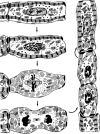
Suggested scheme of myofibril changes throughout mitosis and cell division of adult mammalian cardiomyocytes in vivo. The myofibrils remain unchanged up to late prophase. After the metaphase plate has formed almost all Z-disks are degraded and the myofibrils are subdivided into isolated sarcomeres and myofilament bundles. Complete myofibril disassembly is only seen in telophase. During cytokinesis, the first signs of gradual restoration of Z-disks interconnecting the previously isolated sarcomeres are observed. These early myofibrils are often in disorder, crossing one another. Throughout the subsequent growth and maturation myofibrils become rearranged and oriented parallel to the long axis of the cell. This Figure was published in Rumyantsev, P.P. (1977). Interrelations of the proliferation and differentiation processes during cardiact myogenesis and regeneration. Int Rev Cytol 51,186–273. Copyright Elsevier (1977).
Figure 3
Figure 3
Examples of the problem of interpreting H3P staining in cultured adult cardiomyocytes. (A, C) The H3P-positive nuclei (black) appear to be mitotic cardiomyocyte nuclei indicating proliferation. However, DNA stain (DAPI, black, B, D) reveals in each cell two H3P-negative nuclei, which argue against proliferation as does the presence of intact myofibrils. Possibly, the H3P-positive nuclei belong to overlaying non-myocytes explaining the membrane separating the chromosomes from the cardiomyocytes. Another explanation for C, D is that the membrane is a nuclear membrane separating an endomi-totic cardiomyocyte nucleus.
Figure 4
Figure 4
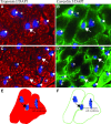
Examples of the problem of identifying cardiomyocyte nuclei in transversal tissue sections of murine myocardium (A–D, 5 μm-thick). Cardiomyocyte-specific cytoplasmic markers (Troponin I, red) are often used to determine the identity of nuclei (DAPI, blue). Cardiomyocyte nuclei (asterices) are embedded in cardiomyocyte cytoplasm whereas non-myocyte nuclei (dots) are not. A muscle-specific membrane marker (Caveolin 3, green), however, shows that some of the embedded nuclei are separated from the cardiomyocyte cytoplasm by a muscle-membrane (arrows). In addition, these nuclei are not surrounded by a continuous muscle membrane. This indicates that these nuclei belong to non-myocytes located in between two cardiomyocytes. The drawings give a schematic overview of this phenomenon (E and F). In case, that this nucleus would indeed belong to a cardiomyocyte one should see a double muscle-membrane.

References

    1. Rosamond W, Flegal K, Friday G, Furie K, Go A, Greenlund K, Haase N, Ho M, Howard V, Kissela B, Kittner S, Lloyd-Jones D, McDermott M, Meigs J, Moy C, Nichol G, O'Donnell CJ, Roger V, Rumsfeld J, Sorlle P, Stelnberger J, Thorn T. Wasserthlel-Smoller S. Hong Y. Heart disease and stroke statistics–2007 update: a report from the American Heart Association Statistics Committee and Stroke Statistics Subcommittee. Circulation. 2007;115:e69–171. - PubMed
    1. Mummery CL. Cardiology: solace for the broken-hearted? Nature. 2005;433:585–7. - PubMed
    1. Rumyantsev PP. Interrelations of the proliferation and differentiation processes during cardiact myogenesis and regeneration. Int Rev Cytol. 1977;51:186–273. - PubMed
    1. Pasumarthi KB, Field LJ. Cardiomyocyte cell cycle regulation. Circ Res. 2002;90:1044–54. - PubMed
    1. Katz AM. Heart failure: a hemodynamic disorder complicated by maladaptive proliferative responses. J Cell Mol Med. 2003;7:1–10. - PMC - PubMed

Publication types