

Review
doi: 10.1111/j.1750-3639.2008.00195.x.
Epub 2008 Jul 25.
Brain extracellular matrix in neurodegeneration
Affiliations
- PMID: 18662234
- PMCID: PMC2742568
- DOI: 10.1111/j.1750-3639.2008.00195.x
Item in Clipboard
Review
Brain extracellular matrix in neurodegeneration
Brain Pathol.
2009 Oct.
Abstract
The role of extracellular matrix (ECM) in neurological development, function and degeneration has evolved from a simplistic physical adhesion to a system of intricate cellular signaling. While most cells require ECM adhesion to survive, it is now clear that differentiated function is intimately dependent upon cellular interaction with the ECM. Therefore, it is not surprising that the ECM is increasingly found to be involved in the enigmatic process of neurodegeneration. Descriptive studies of human neurodegenerative disorders and experimental studies of animal models of neurodegeneration have begun to define potential mechanisms of ECM disruption that can lead to synaptic and neuronal loss.
Figures
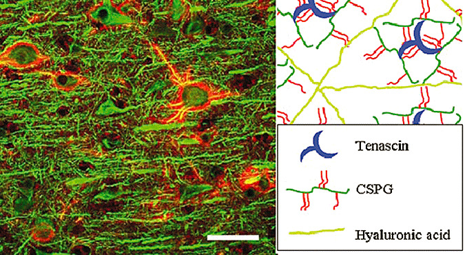
Perineuronal nets (PNNs). Macaque brain stained with microtubule‐associated protein 2 (MAP2) (green) and Wisteria floribunda agglutinin (WFA) (red) (A). Model of PNNs (B). Hypothetical PNN ternary complex of tenascin, chondroitin sulfate proteoglycans (CSPGs) and hyaluronic acid (HA).
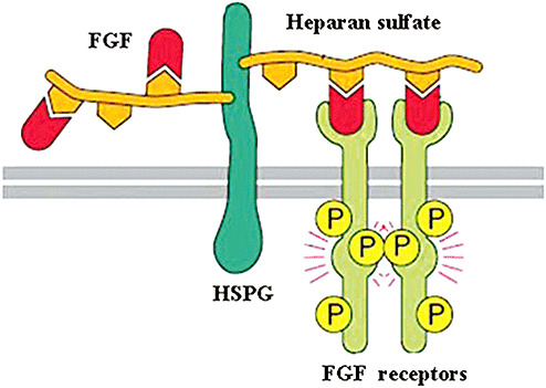
Interaction of heparan sulfate proteoglycans (HSPGs) and fibroblast growth factor (FGF) signaling.
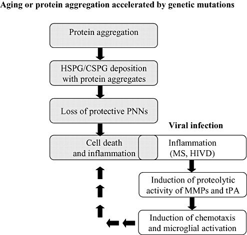
Possible mechanisms of extracellular matrix (ECM)‐related neurodegeneration. Aging or protein aggregation accelerated by genetic mutation can be associated with ECM alterations that would result to co‐deposition of ECM components [eg, heparan sulfate proteoglycans (HSPGs) and chondroitin sulfate proteoglycans (CSPGs)]. Those ECM alterations can result in loss of protective perineuronal nets (PNNs) and increased susceptibility to cell death. Dying neurons can induce inflammation, degradation of ECM and induction of a more robust inflammatory response. Alternatively, inflammatory‐induced neurodegeneration can induce ECM degradation through proteolytic activity [eg, matrix metalloproteinase (MMPs) and tissue plasminogen activator (tPA)], induction of chemotaxis and microglial activation. The resulting trafficking of inflammatory cells and secretion of cytokines can induce neuronal death that would feed the vicious cycle. Abbreviations: MS = multiple sclerosis; HIVD = human immunodeficiency virus dementia.
References
-
- Achim CL, Wiley CA (1996) Inflammation in AIDS and the role of the macrophage in brain pathology. Curr Opin Neurol 9:221–225. - PubMed
-
- Al'Qteishat A, Gaffney J, Krupinski J, Rubio F, West D, Kumar S et al (2006) Changes in hyaluronan production and metabolism following ischaemic stroke in man. Brain 129:2158–2176. - PubMed
-
- Avolio C, Ruggieri M, Giuliani F, Liuzzi GM, Leante R, Riccio P et al (2003) Serum MMP‐2 and MMP‐9 are elevated in different multiple sclerosis subtypes. J Neuroimmunol 136:46–53. - PubMed
Publication types
MeSH terms
Substances
Grants and funding
LinkOut - more resources
Full Text Sources
Other Literature Sources
