Review
. 2008 Jul 25;134(2):215-30.
doi: 10.1016/j.cell.2008.07.001.

TGFbeta in Cancer

Affiliations
Review

TGFbeta in Cancer

Joan Massagué. Cell. .

Abstract

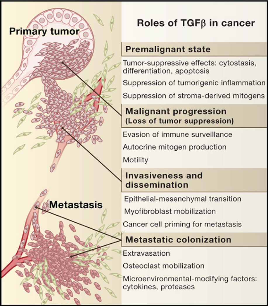
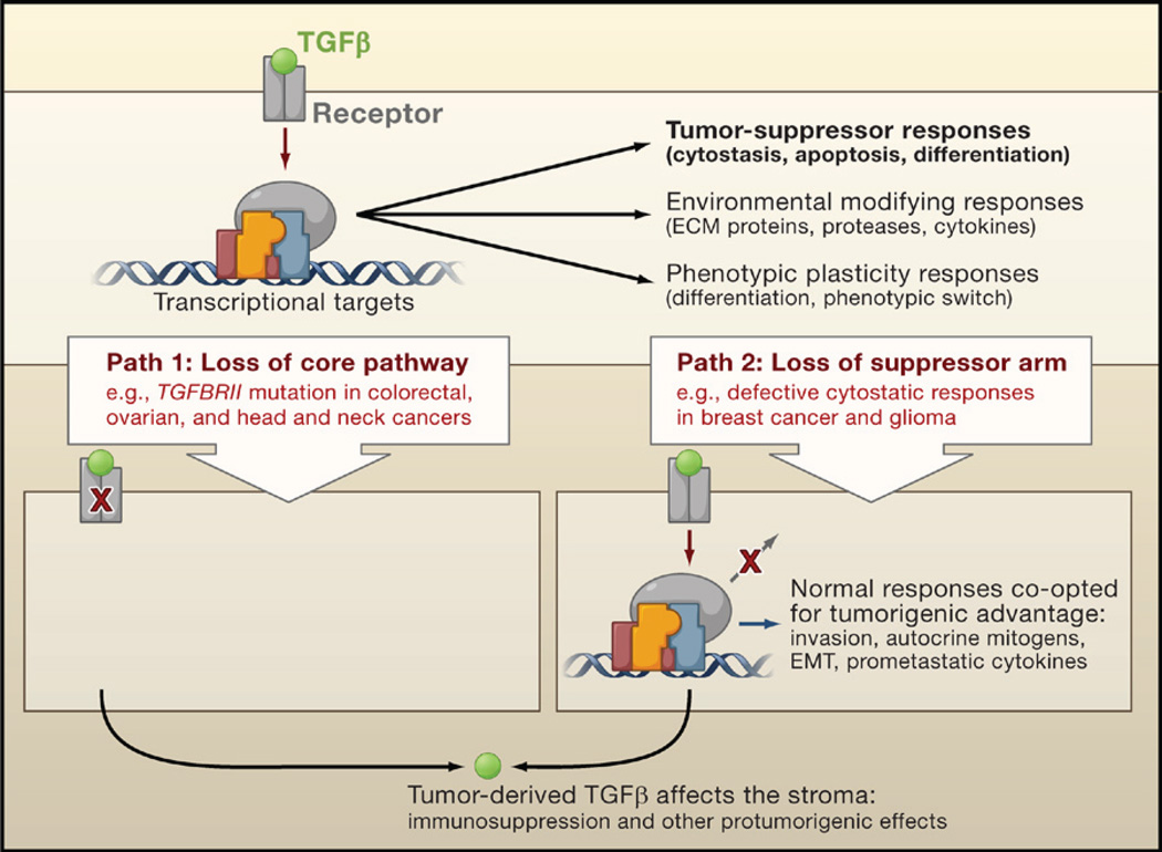
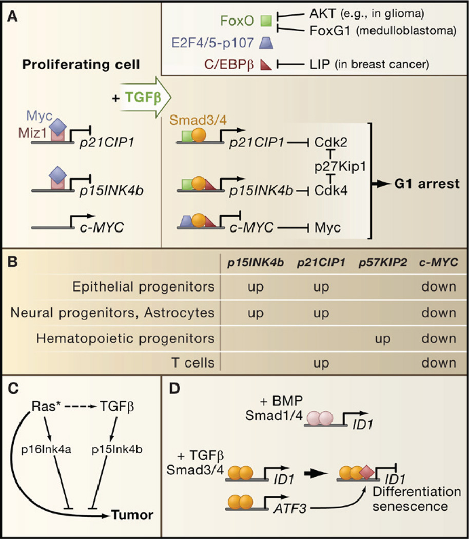
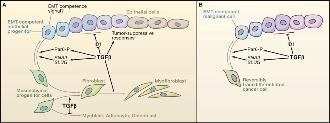
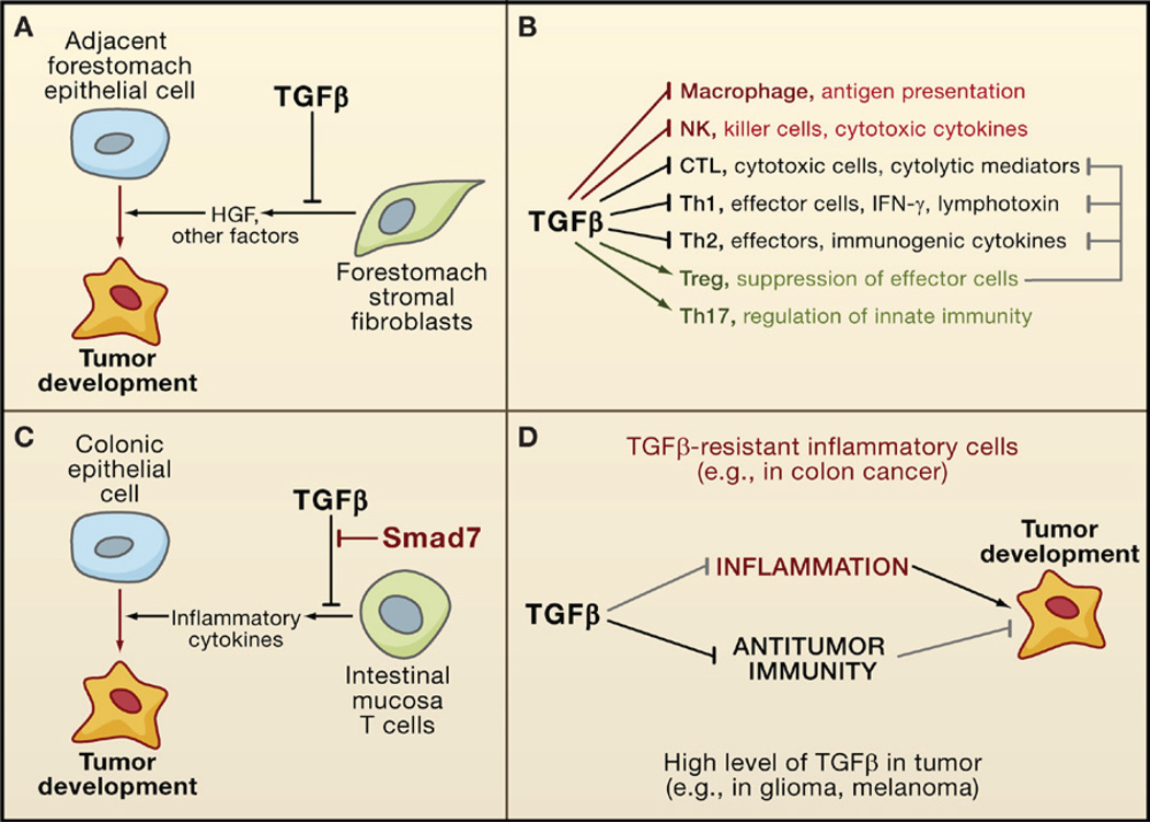
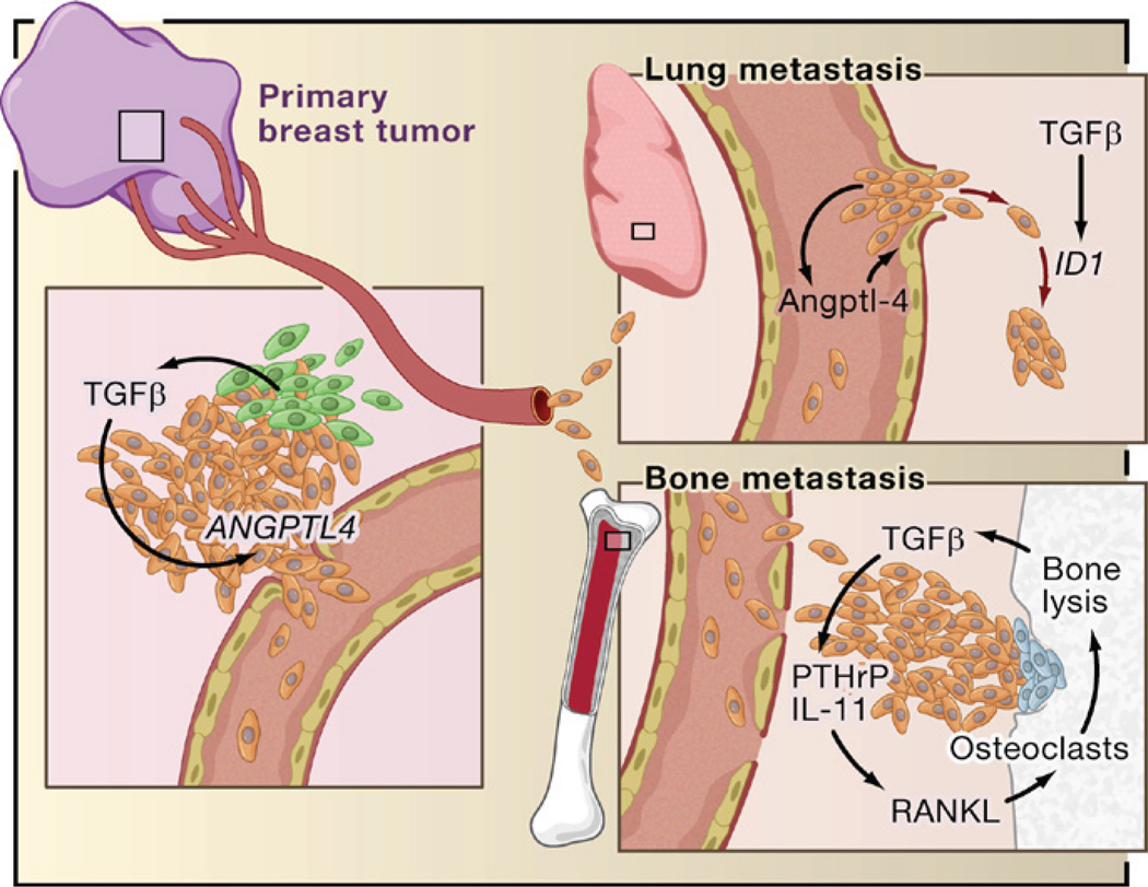
The transforming growth factor beta (TGFbeta) signaling pathway is a key player in metazoan biology, and its misregulation can result in tumor development. The regulatory cytokine TGFbeta exerts tumor-suppressive effects that cancer cells must elude for malignant evolution. Yet, paradoxically, TGFbeta also modulates processes such as cell invasion, immune regulation, and microenvironment modification that cancer cells may exploit to their advantage. Consequently, the output of a TGFbeta response is highly contextual throughout development, across different tissues, and also in cancer. The mechanistic basis and clinical relevance of TGFbeta's role in cancer is becoming increasingly clear, paving the way for a better understanding of the complexity and therapeutic potential of this pathway.


Figures

Figure 1
Figure 1. The Role of TGFβ in Cancer
In normal and premalignant cells, TGFβ enforces homeostasis and suppresses tumor progression directly through cell-autonomous tumor-suppressive effects (cytostasis, differentiation, apoptosis) or indirectly through effects on the stroma (suppression of inflammation and stroma-derived mitogens). However, when cancer cells lose TGFβ tumor-suppressive responses, they can use TGFβ to their advantage to initiate immune evasion, growth factor production, differentiation into an invasive phenotype, and metastatic dissemination or to establish and expand metastatic colonies.
Figure 2
Figure 2. TGFβ and Tumor Progression
TGFβ induces tumor-suppressive effects that cancer cells must circumvent in order to develop into malignancies. Cancer cells can take two alternative paths to this end: (1) decapitate the pathway with receptor-inactivating mutations or (2) selectively amputate the tumor-suppressive arm of the pathway. The latter path allows cancer cells to extract additional benefits by co-opting the TGFβ response for protumorigenic purposes. In both cases, cancer cells can use TGFβ to modulate the microenvironment to avert immune surveillance or to induce the production of protumorigenic cytokines.
Figure 3
Figure 3. Organization of TGFβ Signaling and Weak Links in Cancer
(A) Ligand traps and coreceptor molecules control the access of TGFβ family ligands to signaling receptors. The ligand assembles a tetrameric complex of receptor serine/threonine kinases types I and II. Receptor-II phosphorylates and activates receptor-I, which then phosphorylates and activates Smad transcription factors (RSmads). Activated RSmads bind Smad4 and further build transcriptional activation and repression complexes to control the expression of hundreds of target genes in a given cell. Mitogen-activated protein kinases (MAPK) and other protein kinases phosphorylate Smads for recognition by ubiquitin ligases and other mechanisms of inactivation. Phosphatases have been identified that reverse these phosphorylation events. (B) An abridged chart of ligand-receptor-coreceptor-Smad relationships in the TGFβ (green) and BMP (blue) branches of the TGFβ family. (C) Distinct combinations of transcription partner cofactors in different contexts (e.g., different cell types or conditions) determine the set of genes targeted by specific activated Smads. Each Smad-cofactor combination coordinately regulates a synexpression group of target genes. Smad signaling serves as a node for integrating regulatory signals that impinge on partner cofactors (e.g., Activator signal in Context 2). (D) Alternative modes of TGFβ signaling include Smad4-independent RSmad signaling (via interactions with TIF1γ, IKKα, p68DROSHA), Smad-independent receptor-I signaling (via small G proteins and MAPK pathways), and direct receptor-II signaling (via Par6, and via LIMK1 in the case of BMPR-II). (E) Core TGFβ pathway components that are affected by mutation (red), overexpression (black), or downregulation (green) in human cancers.
Figure 4
Figure 4. Blocking Premalignant Progression by Tumor-Suppressor Proteins
(A) TGFβ and BMP suppress the progression of premalignant states in mouse models. Genetic ablation of TGFβ or BMP receptor genes (TGFBRII and BMPR1A, respectively) or SMAD4 alone does not normally lead to carcinoma formation. However, inactivation of these pathways allows carcinoma progression in transitional epithelia and in premalignant lesions caused by oncogene (KRAS) activation or tumor-suppressor gene (APC) inactivation. (B) Influence of the context on choice of TGFβ tumor-suppressor response. Cells under normal conditions are generally wired for cytostatic or differentiation responses to TGFβ; a loss of TGFβ signaling in this context causes elevated but still regulated cell proliferation (hyperplasia). In contrast, premalignant cells and other hyperproliferative cell states are wired for apoptotic and senescence responses; a loss of TGFβ signaling in this context enables tumor progression (neoplasia).
Figure 5
Figure 5. Tumor-Suppressive Transcriptional Responses to TGFβ
(A) A TGFβ-activated Smad complex in epithelial cells represses c-MYC expression (right panel) and facilitates the induction of CDK inhibitory genes (left panel). Smad-FoxO complexes target p15INK4b and p21CIP1 for transcriptional induction, leading to CDK inhibition. The resulting surge of p15Ink4b releases p27Kip1 from a latent Cdk4-bound state to inhibit CDKs further. FoxO factors can be inhibited by the antagonistic family member FoxG1 or by Akt-mediated phosphorylation in tumors with a hyperactive PI3K-Akt pathway. Overexpression of the C/EBPβ isoform LIP in metastatic breast cancer inhibits C/EBPβ, a common partner of c-MYC and p15INK5b regulatory Smad complexes. (B) Different cell types engage different CDK inhibitor in their TGFβ cytostatic response, whereas c-MYC downregulation is a general feature of the response. (C) p16INK4a induction by endogenous sensors of hyperactive Ras (or other oncogenic signals) collaborates with p15INK4b to mediate tumor suppression. (D) ID1 repression creates conditions for terminal differentiation and senescence. Differential effects of BMP and TGFβ on ID1 expression are based on the ability of TGFβ-activated Smads to recruit the transcriptional repression factor ATF3 to the ID1 regulatory region. Expression of ATF3 itself is induced by the Smad pathway.
Figure 6
Figure 6. Anti- and Protumorigenic Effects of TGFβ on Cell Differentiation
(A) TGFβ favors epithelial differentiation into less proliferative states partly through the downregulation of Inhibitor of Differentiation/DNA binding 1 (ID1). But because of as yet unknown determinants, epithelial progenitor cells can instead become competent to undergo epithelial-mesenchymal transition (EMT) in response to TGFβ. TGFβ functions through the transcription factors SNAIL and SLUG and through phosphorylation of the cell-cell contact regulator Par6 to stimulate EMT. TGFβ also stimulates the differentiation of mesenchymal progenitor cells toward fibroblast and myofibroblast lineages, at the expense of adipocyte and musculoskeletal lineages. (B) Carcinoma cells may avert differentiation into a less proliferative state by switching the ID1 response to TGFβ from repression to activation, as observed in breast cancer cells. Carcinoma progenitor cells that are competent to undergo EMT in response to TGFβ yield highly motile, invasive mesenchymal derivatives, whose presence in tumors is associated with metastatic dissemination.
Figure 7
Figure 7. Anti- and Protumorigenic Effects of TGFβ in the Stroma
(A) TGFβ suppresses tumor emergence in certain epithelial tissues (e.g., the forestomach epithelium) by inhibiting the production of cell survival and motility factors such as hepatocyte growth factor (HGF). (B) TGFβ acts as a major enforcer of immune tolerance by inhibiting the development and functions of nearly all major components of the innate (red) and adaptive (black) immune system. Some of these effects are exerted through the activation of regulatory T cells (Treg; green) that constrain the function of other lymphocytes (gray). (C) By imposing limits on the inflammatory response, TGFβ can avert the protumorigenic effect that could derive from chronic inflammation, as observed in colonic epithelial cells. However, T cells in some patients with inflammatory bowel disease (a colon cancer-prone condition) overexpress Smad7 and are not sensitive to TGFβ. (D) In some types of cancer, a defective TGFβ response in inflammatory cells can lead to excessive inflammation, favoring tumor progression. In other types of cancer, tumor-derived TGFβ can suppress antitumor immune responses, which also favors tumor progression.
Figure 8
Figure 8. Roles of TGFβ in Breast Cancer Metastasis
Based on recent reports, TGFβ derived from infiltrating mesenchymal or myeloid precursor cells (green) or from the cancer cells themselves (brown) in ER breast tumors induces the expression of genes including Angiopoietin-like 4 (ANGPTL4; primary breast tumor inset). Cancer cells entering the circulation with elevated Angptl4 production have an advantage in seeding lung metastasis because of this cytokine’s ability to disrupt vascular endothelial junctions when the cells lodge in lung capillaries (lung metastasis inset). After entering the pulmonary parenchyma, ER breast cancer cells may respond to local TGFβ with induction of Inhibitor of Differentiation/DNA binding 1 (ID1), which acts in this context as a tumor-reinitiating gene. The entry of circulating tumor cells into the bone marrow does not benefit from Angptl4 because these capillaries are naturally fenestrated to allow the constant passage of cells (bone metastasis inset). However, TGFβ released by osteoclasts (blue) from rich stores in the bone matrix acts on the growing cancer cells to stimulated the production of parathyroid hormone-related protein (PTHrP) and interleukin- 11. These factors act on osteoblasts to release RANK ligand (RANKL) and other mediators of osteoclast moblization, perpetuating the osteolytic metastasis cycle.

References

    1. Allinen M, Beroukhim R, Cai L, Brennan C, Lahti-Domenici J, Huang H, Porter D, Hu M, Chin L, Richardson A, et al. Molecular characterization of the tumor microenvironment in breast cancer. Cancer Cell. 2004;6:17–32. - PubMed
    1. Arteaga CL. Inhibition of TGFbeta signaling in cancer therapy. Curr. Opin. Genet. Dev. 2006;16:30–37. - PubMed
    1. Ashcroft GS, Yang X, Glick AB, Weinstein M, Letterio JL, Mizel DE, Anzano M, Greenwell-Wild T, Wahl SM, Deng C, Roberts AB. Mice lacking Smad3 show accelerated wound healing and an impaired local inflammatory response. Nat. Cell Biol. 1999;1:260–266. - PubMed
    1. Bardeesy N, Cheng KH, Berger JH, Chu GC, Pahler J, Olson P, Hezel AF, Horner J, Lauwers GY, Hanahan D, DePinho RA. Smad4 is dispensable for normal pancreas development yet critical in progression and tumor biology of pancreas cancer. Genes Dev. 2006;20:3130–3146. - PMC - PubMed
    1. Becker C, Fantini MC, Neurath MF. TGF-beta as a T cell regulator in colitis and colon cancer. Cytokine Growth Factor Rev. 2006;17:97–106. - PubMed

Publication types

MeSH terms

Substances

LinkOut - more resources