. 2008 Jul 25;134(2):256-67.
doi: 10.1016/j.cell.2008.05.043.

The Cdc14B-Cdh1-Plk1 axis controls the G2 DNA-damage-response checkpoint

Affiliations

The Cdc14B-Cdh1-Plk1 axis controls the G2 DNA-damage-response checkpoint

Florian Bassermann et al. Cell. .

Abstract

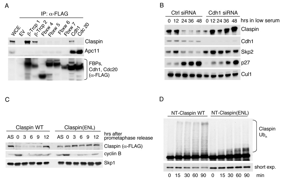
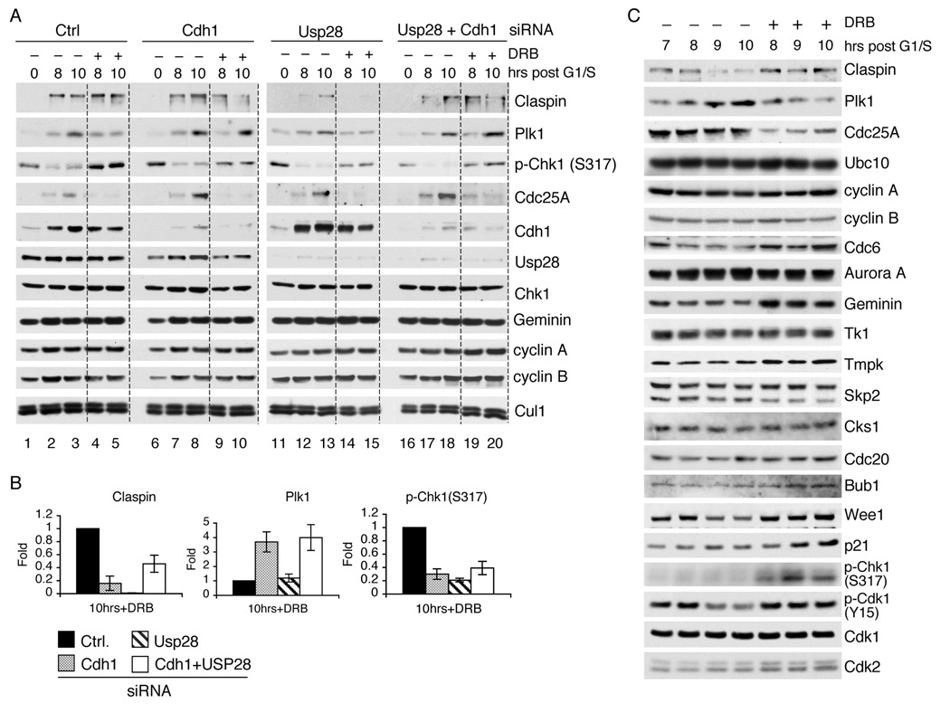
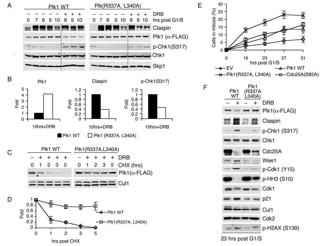
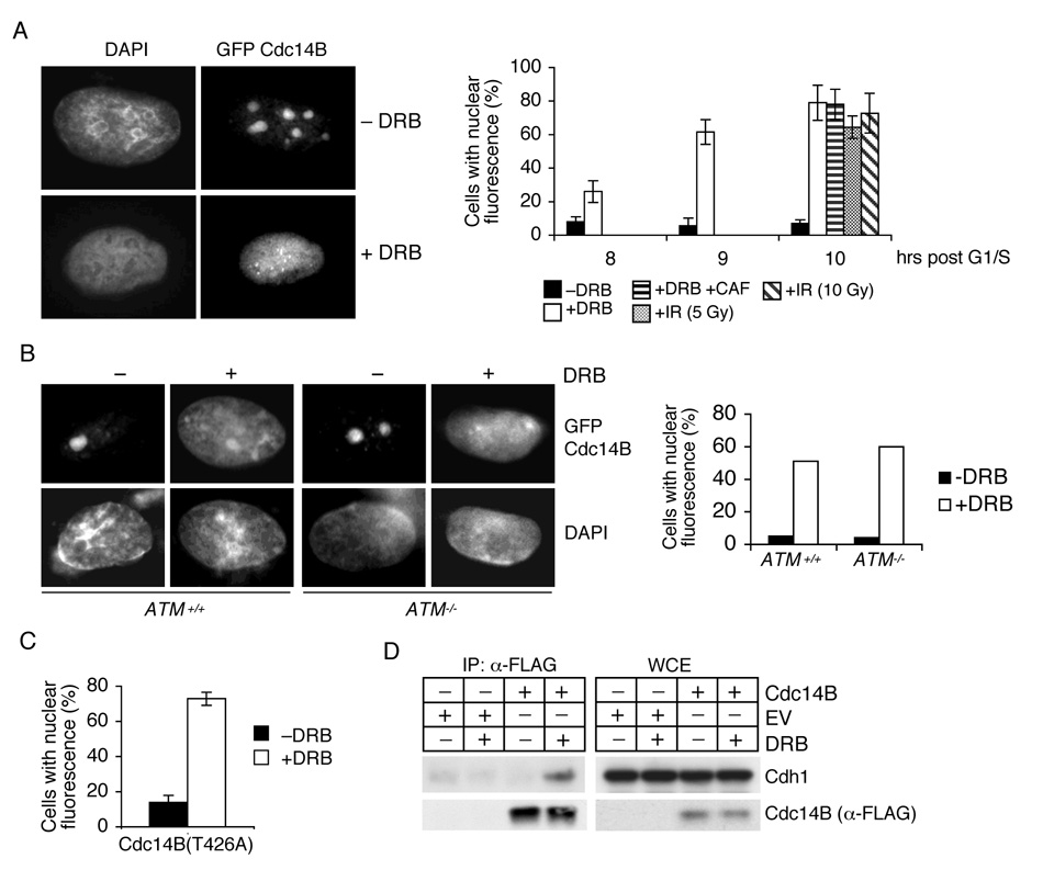
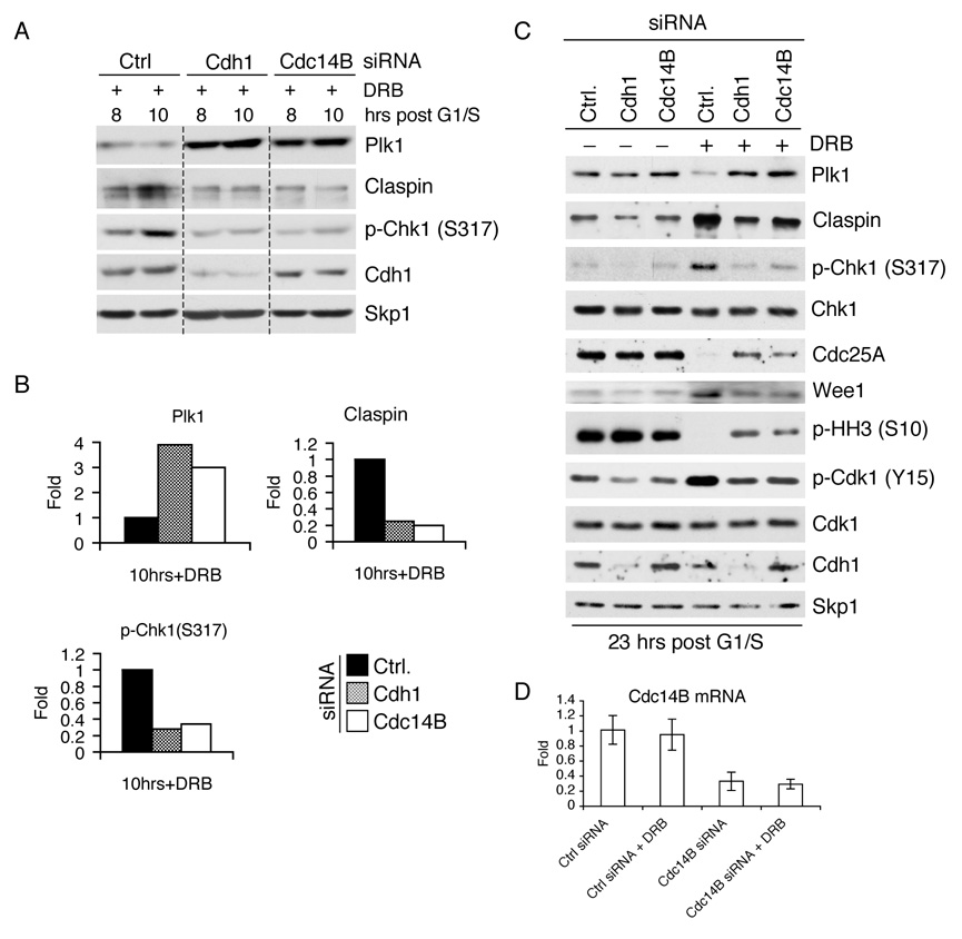
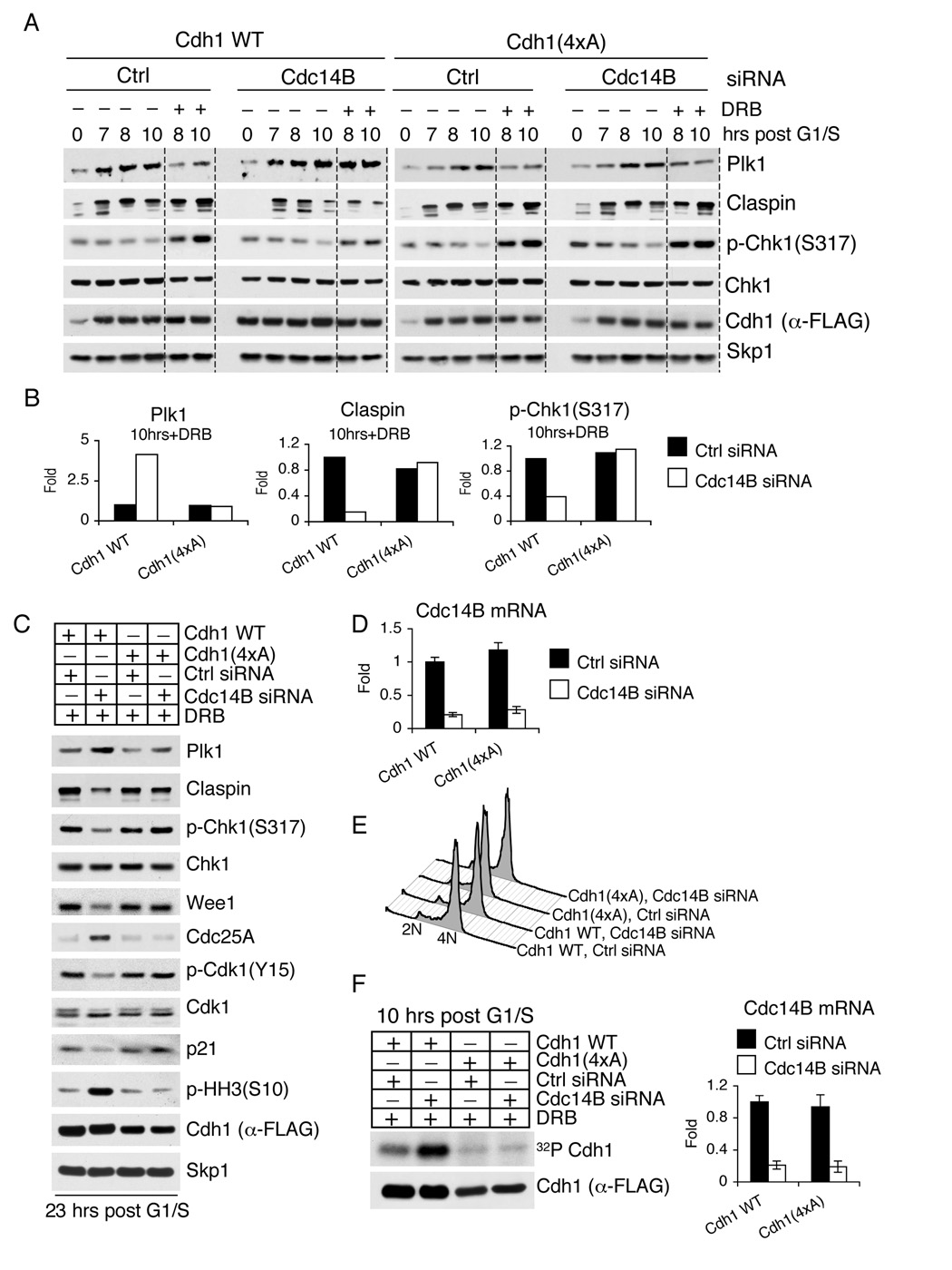
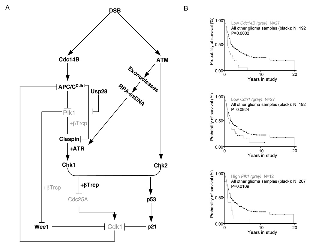
In response to DNA damage in G2, mammalian cells must avoid entry into mitosis and instead initiate DNA repair. Here, we show that, in response to genotoxic stress in G2, the phosphatase Cdc14B translocates from the nucleolus to the nucleoplasm and induces the activation of the ubiquitin ligase APC/C(Cdh1), with the consequent degradation of Plk1, a prominent mitotic kinase. This process induces the stabilization of Claspin, an activator of the DNA-damage checkpoint, and Wee1, an inhibitor of cell-cycle progression, and allows an efficient G2 checkpoint. As a by-product of APC/C(Cdh1) reactivation in DNA-damaged G2 cells, Claspin, which we show to be an APC/C(Cdh1) substrate in G1, is targeted for degradation. However, this process is counteracted by the deubiquitylating enzyme Usp28 to permit Claspin-mediated activation of Chk1 in response to DNA damage. These findings define a novel pathway that is crucial for the G2 DNA-damage-response checkpoint.


Figures

Figure 1
Figure 1. Claspin is degraded in G0 and G1 via the APC/CCdh1 ubiquitin ligase
(A) Cdh1 interacts with Claspinin vivo HEK293T cells were transfected with the indicated FLAG-tagged constructs or an empty vector (EV). Whole cell extracts (WCE) were immunoprecipitated (IP) with anti-FLAG resin, and immunocomplexes were probed with antibodies to the indicated proteins. (B) T98G cells were transfected with a control (Ctrl) siRNA oligo or an siRNA oligo directed against Cdh1 mRNA. Cells were then switched to culture media containing 0.02% FBS to arrest them in G0/G1. Samples were collected at the indicated times after the beginning of the serum starvation and subjected to immunoblot analysis using antibodies to the indicated proteins. Skp2 and p27 were used as cell cycle markers. (C) Claspin(ENL) is stable in G1. HeLa cells were infected with retroviruses expressing either FLAG-tagged wild type Claspin or FLAG-tagged Claspin(ENL) and subsequently treated for 16 hours with nocodazole to induce a mitotic block. Round, prometaphase cells were then collected by gentle shake-off and replated in fresh medium for the indicated times. Cells were harvested, and cell extracts were analyzed by immunoblotting with antibodies to the indicated proteins. Synchronization was monitored by flow cytometry and by the levels of cyclin B. (D) Ubiquitin ligation assays of 35S-labeled, in vitro translated Claspin N-terminal fragments (amino acid 1–678) of either wild type Claspin or Claspin(ENL) were conducted in the presence of unlabeled, in vitro translated Cdh1. Samples were incubated at 30°C for the indicated times. The lower autoradiography image represents a short exposure time, and the upper image represents a long exposure time. The bracket on the right side of the top panel marks a ladder of bands corresponding to polyubiquitylated Claspin.
Figure 2
Figure 2. In response to DNA damage in G2, APC/CCdh1 is reactivated to target Plk1 and Claspin (but the latter is protected by Usp28)
(A) U2OS cells were transfected with the indicated siRNA oligos and synchronized at G1/S using a double thymidine block. Cells were then released from the block to allow progression towards G2. At seven hours post release, cells were pulsed for one hour with either solvent (−) or doxorubicin (DRB) (+) and collected at the indicated times thereafter. Whole cell extracts were analyzed by immunoblotting with antibodies to the indicated proteins. Synchrony was verified by flow cytometry. To facilitate comparison, a gray line separates samples treated with doxorubicin from untreated samples. (B) The graphs show the quantification of the levels of Claspin, Plk1, and Chk1 phosphorylated on Ser317 shown in (A) at the 10 hour timepoint averaged with two additional, independent experiments. The value given for the amount of protein present in the control sample two hours after the end of the doxorubicin pulse was set as 1 (n=3, ± SD). (C) U2OS cells were synchronized and treated with DRB as described in (A). Samples were collected at the indicated times and processed for immunoblot analysis with antibodies to the indicated proteins.
Figure 3
Figure 3. The DNA damage checkpoint in G2 requires Cdh1-dependent degradation of Plk1
(A) U2OS cells retrovirally infected with the indicated Plk1 constructs were synchronized and treated with DRB as described in (2A). Cells were then collected at the indicated times and immunoblotted with antibodies to the indicated proteins. To facilitate comparison, a gray line separates samples treated with doxorubicin from untreated samples. (B) The graphs show the quantification of the levels of Claspin, Plk1, and Chk1 phosphorylated on Ser317 shown in (A) at the 10 hour timepoint averaged with an additional, independent experiment. The value given for the amount of protein present in the control sample two hours after the end of the doxorubicin pulse was set as 1 (n=2). (C) U2OS cells were infected, synchronized, and pulsed with doxorubicin as in (A), except that cycloheximide (CHX) was added seven hours after release from G1/S. At different times after the addition of CHX, cells were collected, and cell lysates were immunoblotted with antibodies to the indicated proteins. (D) The graph shows the quantification of the levels of wild type Plk1 and Plk1(R337A,L340A) shown in (C) averaged with two additional, independent experiments (n=3, ± SD). (E) U2OS cells infected with either an empty virus (EV) or retroviruses encoding wild type Plk1, Plk1(R337A,L340A), or Cdc25A(S82A) were synchronized and treated with DRB as described in (A). Thereafter, cells were incubated in fresh medium containing nocodazole to trap cells in mitosis. Samples were then collected at the indicated times, and the percentage of mitotic cells was monitored by immunodetection of Histone H3 phosphorylated on Ser10 using flow cytometry (n=3, ± SD). (F) U2OS cells treated as in (A) were collected 23 hours after release from G1/S (16 hours after the doxorubicin pulse), and then cell lysates were immunoblotted with antibodies to the indicated proteins.
Figure 4
Figure 4. Cdh1 associates with Cdc14B in G2 in response to DNA damage
(A) Cdc14B moves from the nucleolus to the nucleoplasm in response to DNA damage. U2OS cells transfected with a construct expressing GFP-tagged Cdc14B were synchronized as described in (2A) and then either treated with DRB [+/−caffeine (CAF)] or subjected to ionizing radiations (IR). Cells were then collected and analyzed by direct immunofluorescence. Left panels: micrographs of representative cells showing the subcellular localization of Cdc14B. Right panel: Quantification of cells with nuclear Cdc14B fluorescence at the indicated times post pulse with doxorubicin (n=3, ± SD). (B) Cdc14B translocation to the nucleus is independent of ATM. The experiment was performed as in (A), except that asynchronous ATM+/+ and ATM−/− fibroblasts were used (n=2). (C) The experiment was performed as in (A), except that GFP-tagged Cdc14B(T426A) was used (n=3). (D) Cdh1 binds to Cdc14B in a DNA damage-dependent manner. U2OS cells infected with either an empty retrovirus (EV) or viruses encoding FLAG-tagged Cdc14B were synchronized and treated with DRB as described in (A). Cells were collected two hours later, and whole cell extracts (WCE) were immunoprecipitated (IP) with anti-FLAG resin. The indicated proteins were detected by immunoblotting.
Figure 5
Figure 5. DNA damage-dependent reactivation of APC/CCdh1 in G2 requires Cdc14B
(A) U2OS cells transfected with the indicated siRNA oligos were synchronized and treated with DRB as described in (2A). Cells were collected at the indicated times, and cell lysates were analyzed by immunoblotting with antibodies to the indicated proteins. (B) The graphs show the quantification of Claspin, Plk1, and Chk1 phosphorylated on Ser317 shown in (A) at the 10 hour timepoint averaged with an additional, independent experiment. The value given for the amount of protein present in the control sample two hours after the end of the doxorubicin pulse was set as 1 (n=2). (C) U2OS cells, transfected with the indicated siRNA oligos, were treated as in (A). Twenty-three hours after release from G1/S (16 hours after the doxorubicin pulse), cells were collected, and cell lysates were analyzed by immunoblotting with antibodies to the indicated proteins. (D)Cdc14B mRNA levels of cells used in (A) and (C) were analyzed eight hours after release from G1/S using real time PCR in triplicate measurements (± SD). The value given for the amount of Cdc14B mRNA present in the sample treated with control oligos was set as 1.
Figure 6
Figure 6. A constitutively active Cdh1 mutant is refractory to the silencing of Cdc14B
(A) U2OS cells were retrovirally infected with either wild type Cdh1 or Cdh1(4xA) mutant and subsequently transfected with the indicated siRNA oligos. Cells were then synchronized and treated with DRB as described in (2A), collected at the indicated times, and immunoblotted with antibodies to the indicated proteins. To facilitate comparison, a gray line separates samples treated with doxorubicin from untreated samples. (B) The graphs show the quantification of the levels of Claspin, Plk1, and Chk1 phosphorylated on Ser317 shown in (A) at the indicated timepoint averaged with an additional, independent experiment. The value given for the amount of protein present in the control sample two hours after the end of the doxorubicin pulse was set as 1 (n=2). (C) U2OS cells expressing wild type Cdh1 or Cdh1(4xA) were transfected with the indicated siRNA oligos and treated as in (A). Twenty-three hours after release from G1/S (16 hours after the doxorubicin pulse), cells were collected, and cell lysates were analyzed by immunoblotting with antibodies to the indicated proteins. (D)Cdc14B mRNA levels of cells used in (A) and (C) were analyzed eight hours after release from G1/S using real time PCR in triplicate measurements (± SD). The value given for the amount of Cdc14B mRNA present in the sample expressing wild type Cdh1 and treated with control oligos was set as 1. (E) Synchrony in G2 was ascertained by flow cytometry for cells used in (A) at the time of doxorubicin treatment. Identical synchrony in G2 was obtained for cells used in (C). (F) The experiment was performed as in (A), except that an in vivo labeling with 32P-orthophosphate was performed during the last three hours before cells were collected. Cdh1 was then immunoprecipitated under denaturing conditions, resolved by SDS-PAGE, and visualized by autoradiography (upper panel) or immunoblotting (bottom panel). Right panel shows Cdc14B mRNA levels analyzed by real time PCR.
Figure 7
Figure 7. The Cdc14B-Plk1-Cdh1 axis controls the DNA damage response in G2 and is deregulated in human tumors
(A) A model of the G2 DNA damage response checkpoint. Black signifies activated forms of the respective proteins, and gray indicates inactive forms or degraded proteins. After induction of double strand breaks (DSB) in G2, ATM activates Chk2. In addition, ATM activates certain exonucleases to induce DSB resection, resulting in RPA coated, single-stranded DNA (ssDNA), which contributes to the recruitment of ATR. In parallel, Cdc14B is released from the nucleolus to the nucleoplasm, activating APC/CCdh1, which in turn targets Plk1 for proteasomal degradation. Because of the low levels of Plk1, phosphorylation of Claspin and Wee1 is reduced, preventing βTrcp-mediated degradation. Claspin is protected from APC/CCdh1-mediated degradation by Usp28. Stable Claspin promotes the ATR-mediated activation of Chk1, which, together with Chk2, targets Cdc25A (inducing its degradation) and p53 (promoting its stabilization and consequent induction of p21). As a result, Cdk1 activity is attenuated, and cells arrest in G2. Stable Wee1 contributes to this inhibition by directly phosphorylating Cdk1. The reduction in Cdk1 activity further removes the constraints on APC/CCdh1 activity. (B) Poor patient survival correlates with low levels of Cdc14B and Cdh1 and high levels of Plk1 (gray).Rembrandt glioma database (http://rembrandt.nci.nih.gov)-generated Kaplan-Meier survival curves of 219 patients with gliomas of all histological grades grouped by gene expression levels of Cdc14B (top plot), Cdh1 (middle plot), and Plk1 (bottom plot). The associated P values are shown for each plot.

Comment in

References

    1. Amador V, Ge S, Santamaria P, Guardavaccaro D, Pagano M. APC/C(Cdc20) controls the ubiquitin-mediated degradation of p21 in prometaphase. Mol Cell. 2007;27:462–473. - PMC - PubMed
    1. Bartek J, Lukas J. DNA damage checkpoints: from initiation to recovery or adaptation. Curr Opin Cell Biol. 2007;19:238–245. - PubMed
    1. Bassermann F, von Klitzing C, Munch S, Bai R, Kawaguchi H, Morris W, Peschel C, Duyster J. NIPA defines an SCF-type mammalian E3 ligase that regulates mitotic entry. Cell. 2005;122:45–57. - PubMed
    1. Busino L, Donzelli M, Chiesa M, Guardavaccaro D, Ganoth D, Dorrello N, Hershko A, Pagano M, Draetta GF. Degradation of Cdc25A by αTrCP during S phase and in response to DNA damage. Nature. 2003;426:87–91. - PubMed
    1. Cho P, Liu Y, Gomez M, Dunlap J, Tyers M, Wang Y. The dual-specificity phosphatase CDC14B bundles and stabilizes microtubules. Mol Cell Biol. 2005;25:4541–4551. - PMC - PubMed

Publication types

MeSH terms

Substances