. 2008 Sep 15;337(2):121-31.
doi: 10.1016/j.jim.2008.07.005. Epub 2008 Jul 26.

Optimization of methods to study pulmonary dendritic cell migration reveals distinct capacities of DC subsets to acquire soluble versus particulate antigen

Affiliations

Optimization of methods to study pulmonary dendritic cell migration reveals distinct capacities of DC subsets to acquire soluble versus particulate antigen

Claudia Jakubzick et al. J Immunol Methods. .

Abstract

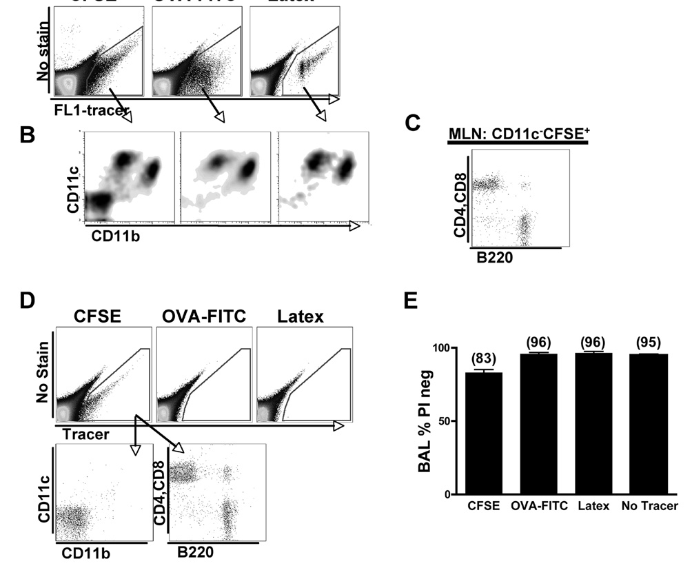
Dendritic cell migration from the airway to lymph nodes is a key event in the development of airway immunity during infection, allergy, and vaccination. To identify the best approaches to investigate DC migration to lung-draining lymph nodes, we directly compared three methods previously used to track DC migration: airway administration of fluorescent OVA, latex beads, or carboxyfluorescein succinimidyl ester (CFSE). We show that two of the methods employed in optimal conditions-administration of fluorescent OVA or latex particles-have broadly relevant utility in studies of pulmonary DC migration, both in the presence and absence of inflammatory mediators. However, CFSE was of limited value because it induced a robust airway inflammatory response upon instillation. Unexpectedly, antigen-loaded tracers with distinct physical properties differently affected the populations that acquired the tracers and the overall T cell response. Specifically, soluble OVA and OVA formulated as a particulate after conjugation to latex beads were acquired in different proportions in vivo by the two characterized subsets of pulmonary DCs: CD11b(hi)CD103(-) and CD11b(lo)CD103(+)langerin(+) DC populations. Consequently, and in line with recent studies that these two subsets of DCs respectively activate CD4(+) and CD8(+) lymphocyte populations, the physical nature of the antigen delivery vehicle strongly influenced the degree of CD4(+) versus CD8(+) OVA-specific T cell activation. This finding suggests that changes in the physical presentation of the same antigen delivered to the airway during natural immune responses or vaccinations may markedly affect the character of the T cell response that ensues.


Figures

Figure 1
Figure 1. Labeling methods compared in BAL and Lung
A) Flow cytometry of BAL gated on live cells 1 day post IN delivery of CFSE, OVA-FITC or latex particles without or with 1µg LPS, stained for CD11c versus the fluorescein fluorescent channel (left). Neutrophils are gated in red in plots depicting CD11c versus CD11b staining. Top panel shows IN delivery of PBS containing no tracer: DCs are gated in green and alveolar macrophages are gated in blue. In addition, the presence of non-latex+ DCs could be observed in the mice that received latex delivery (green gate). Representative data from seven experiments B) Lung histology of labeling methods top row without LPS: tracer is green; MHCII is red; and DAPI is blue. Naïve mouse lung (no tracer) bottom row: MHCII and control antibody are red and DAPI is blue. 200X magnification. C) Flow cytometry of BAL gated on live cells stained for CD11c versus CD11b one-day post IN delivery of commercially available OVA-FITC non-dialyzed and dialyzed. Non-FITC conjugated egg-extracted OVA was used as a control (left). D) Flow cytometry of BAL gated on live cells stained for CD11c versus CD11b one-day post IN delivery of FITC-conjugated, egg-extracted OVA and commercially available OVA. Bar graph measured the number of neutrophil infiltration into the BAL.
Figure 2
Figure 2. Tracer+ pulmonary DCs in the MLN
A) Gated live cells from MLN were plotted as an empty channel versus FITC and then all tracer+ cells were gated. B) Gated MLN tracer+ cells were plotted as CD11c versus CD11b. These mice were treated with 1µg LPS. Data are representative of four experiments. C) CD11cCD11bCFSE+ cells from the MLN were stained for T cell markers, CD4 and CD8, and for a B cell marker, B220. D) Gated live cells from inguinal LN (distal LN) were plotted as an empty channel versus tracer one-day post IN delivery of CFSE, commercial OVA-FITC and latex particles. Lower plots show the CFSE+ cells from the inguinal LN plotted as CD11c versus CD11b and CD4+CD8 versus B220. Data are representative of three independent experiments. E) Analysis of the percent of live cells present in the BAL one-day post IN delivery of labeling methods. The percentage of live cells was analyzed by gating on negatively stained propidium iodide cells: n=5–7 mice per group.
Figure 3
Figure 3. Time course analysis of migrating tracer+ cell in the MLN and the percentage and number of tracer+ cells found in the MLN
A) All live cells are plotted as CD11c versus CD11b and then all CD11chi cells in the MLN were gated, two main subsets are observed: CD11chiCD11blo and CD11chiCD11bhi DCs. Within these subsets, tracer+ migratory DCs were extracted. Next, all CD11chi cells were plotted as an empty channel versus tracer and then all tracer+ cells were gated on (migrating DCs). Small gray arrow in the latex method top panel represents the CD11cintCD11b cells, where the majority of these cells are B220+ plasmacytoid DCs. These mice were treated with 1µg LPS. Data are representative of five independent experiments. B) Gated latex particle+ DCs were stained for CD103 or CD8 versus CD11b. C) CD11c+ BAL DCs were gated by low autofluorescence (macrophages are the highly autofluorescent) using empty FITC fluorescent channel, then the DCs were stained for CD103 or CD8 versus CD11b in naïve and mice treated with 1µg LPS. D) Time course indicating the total number of migrated pulmonary DCs, CD11chiFITC+ cells in the MLN post IN delivery of tracer with LPS E) The percentage and number of tracer+ cells (CFSE+, EE=FITC conjugated egg extracted OVA, commercial OVA-FITC+ and latex particle+) in CD11chi DCs in all labeling methods one-day post IN delivery with or without LPS: n=8–10 mice per group. Results are expressed as mean ±SEM (error bars); difference between EE and commercial OVA-FITC treated mice with or without LPS was significant as was latex particle non-LPS and LPS treated mice (*p<0.05, **p<0.001, ***p<0.0001).
Figure 4
Figure 4. Pertussis toxin significantly inhibited pulmonary DC migration to the MLN
A) Representative dot plots in the BAL were stained for CD11c versus CD11b showing neutrophil infiltration after one-day post IN delivery of tracer with 1µg LPS. In addition, some mice were treated simultaneously with PBS (top plots) or 1µg of pertussis toxin (bottom plots). Bar graph summarizes the percentage of neutrophils present in BAL from PBS or pertussis toxin treated mice one-day post IN delivery of CFSE, commercial OVA-FITC and latex particles: n=5 mice per group. B) Dot plots display gated CFSE+, OVA-FITC+ and latex particle+ cells in the MLN stained for CD11c versus CD11b in PBS and 1µg pertussis toxin treated mice. The bar graph compiles the total number of CD11chitracer+ (CFSE+, OVA-FITC+ and latex particle+) cells in the MLN from LPS labeled mice treated with PBS or pertussis toxin: n=5 mice per group. Results are expressed as mean ±SEM (error bars); differences between PBS and pertussis toxin treated mice were significant for all tracers (**p<0.001, *** p<0.0001).
Figure 5
Figure 5. CD11bhi DCs preferentially acquires soluble protein, whereas CD11bloCD103+ DCs preferentially take up particles
A) The percentage of CD11blo and CD11bhi DCs in CD11chitracer+ cells from IN delivery of OVA-FITC or latex particles: n=8–10 mice per group. Result is expressed as mean ±SEM (error bars); difference between OVA-FITC and latex particle treated mice is statistically significant for the percentage of FITC+CD11blo or FITC+CD11bhi DCs (*** p<0.0001). B) Left bar graph shows the percentage of CD11blo and CD11bhi DCs in CD11chitracer+ cells from IN delivery of CFSE and bar graph on the right shows the percentage of all (tracer+ and tracer) CD11blo and CD11bhi DCs in the MLN from the three types of tracer delivered mice: n=8–10 mice per group.
Figure 6
Figure 6. OT-I cells proliferate more when CD103+ DCs present antigen compared with presentation by CD11bhi DCs
Representative histograms show gated adoptively transferred Vα2+CFSE+ OT-I and OT-II cells in the MLN (top row) for IN delivery of non-conjugated latex particles as a negative control, i.p. injection of commercial OVA as a positive control, IN delivery of egg-extracted OVA-conjugated latex particles and egg-extracted soluble OVA-FITC. Below the first row, Vα2+CFSE+ gate was divided and viewed as a histogram for proliferation of CD8+ OT-I and CD4+ OT-II cells. Bar graph (on the left) demonstrates the ratio of the number of OT-I/OTII cells found in the MLN 4 days after adoptive transfer in mice treated with non-conjugated latex particles. Bar graph (right) the ratio of the number of OT-I/OT-II cells that proliferated in the MLN 4 days after adoptive transfer in mice treated with egg-extracted OVA-conjugated latex particles and egg-extracted OVA-FITC. Data are representative of three independent experiments. Result is expressed as mean ±SEM (error bars); difference between mice treated with EE OVA-conjugated latex particles and EE OVA-FITC is statistically significant (*** p<0.0001).

References

    1. Barnden MJ, Allison J, Heath WR, Carbone FR. Defective TCR expression in transgenic mice constructed using cDNA-based alpha- and beta-chain genes under the control of heterologous regulatory elements. Immunol Cell Biol. 1998;76:34–40. - PubMed
    1. Beaty SR, Rose CE, Jr, Sung SS. Diverse and Potent Chemokine Production by Lung CD11bhigh Dendritic Cells in Homeostasis and in Allergic Lung Inflammation. J Immunol. 2007;178:1882–1895. - PubMed
    1. Belz GT, Bedoui S, Kupresanin F, Carbone FR, Heath WR. Minimal activation of memory CD8+ T cell by tissue-derived dendritic cells favors the stimulation of naive CD8+ T cells. Nat Immunol. 2007;8:1060–1066. - PubMed
    1. Belz GT, Smith CM, Kleinert L, Reading P, Brooks A, Shortman K, Carbone FR, Heath WR. Distinct migrating and nonmigrating dendritic cell populations are involved in MHC class I-restricted antigen presentation after lung infection with virus. Proc Natl Acad Sci U S A. 2004;101:8670–8675. - PMC - PubMed
    1. Brimnes MK, Bonifaz L, Steinman RM, Moran TM. Influenza virus-induced dendritic cell maturation is associated with the induction of strong T cell immunity to a coadministered, normally nonimmunogenic protein. J Exp Med. 2003;198:133–144. - PMC - PubMed

Publication types