. 2008 Jul 28;182(2):263-76.
doi: 10.1083/jcb.200710195.

Extensive meiotic asynapsis in mice antagonises meiotic silencing of unsynapsed chromatin and consequently disrupts meiotic sex chromosome inactivation

Affiliations

Extensive meiotic asynapsis in mice antagonises meiotic silencing of unsynapsed chromatin and consequently disrupts meiotic sex chromosome inactivation

Shantha K Mahadevaiah et al. J Cell Biol. .

Abstract

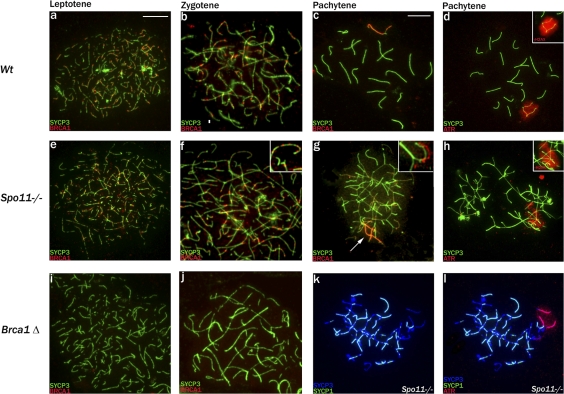
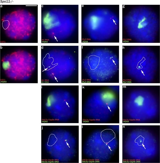
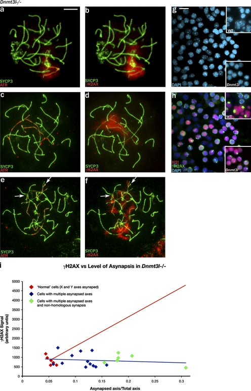
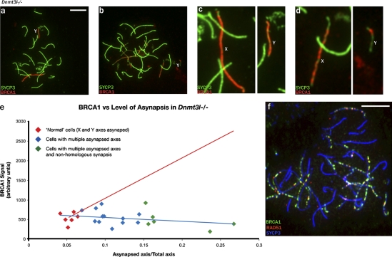
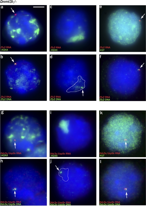
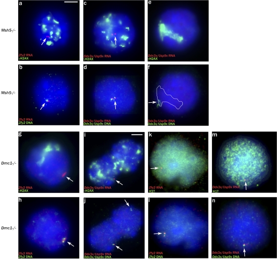
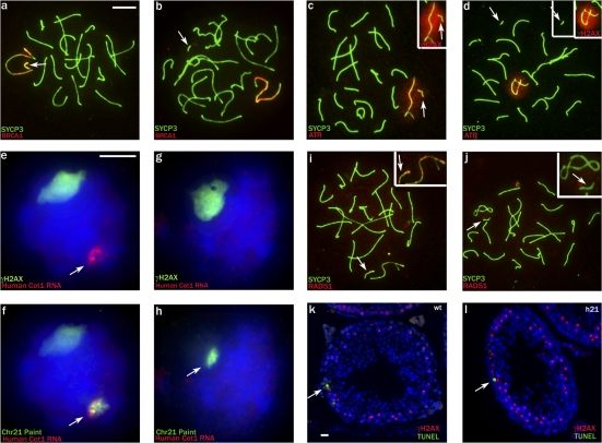
Chromosome synapsis during zygotene is a prerequisite for the timely homologous recombinational repair of meiotic DNA double-strand breaks (DSBs). Unrepaired DSBs are thought to trigger apoptosis during midpachytene of male meiosis if synapsis fails. An early pachytene response to asynapsis is meiotic silencing of unsynapsed chromatin (MSUC), which, in normal males, silences the X and Y chromosomes (meiotic sex chromosome inactivation [MSCI]). In this study, we show that MSUC occurs in Spo11-null mouse spermatocytes with extensive asynapsis but lacking meiotic DSBs. In contrast, three mutants (Dnmt3l, Msh5, and Dmc1) with high levels of asynapsis and numerous persistent unrepaired DSBs have a severely impaired MSUC response. We suggest that MSUC-related proteins, including the MSUC initiator BRCA1, are sequestered at unrepaired DSBs. All four mutants fail to silence the X and Y chromosomes (MSCI failure), which is sufficient to explain the midpachytene apoptosis. Apoptosis does not occur in mice with a single additional asynapsed chromosome with unrepaired meiotic DSBs and no disturbance of MSCI.


Figures

Figure 1.
Figure 1.
The MSUC initiator proteins BRCA1 and ATR are recruited to the pseudo–sex body domain of Spo11-null pachytene spermatocytes. (a–c) SYCP3 and BRCA1 staining of control spermatocytes showing the appearance of BRCA1 foci on the forming axial elements in leptotene, their loss after synapsis during zygotene, and BRCA1 accumulation on the asynapsed X and Y axes in pachytene. (d) The kinase ATR appears throughout the chromatin of the asynapsed X and Y axes in pachytene of control males, and there is an associated phosphorylation of H2AX (γH2AX), which defines the sex body domain (inset). (e–g) In Spo11-null spermatocytes, which lack meiotic DSBs, BRCA1 foci still appear on the axial elements in leptotene, are retained throughout the asynaptic zygotene stage (magnified in insets), and are lost in conjunction with the nonhomologous synapsis apparent in pachytene (highlighted in insets, which show synapsed and asynapsed axes with BRCA1 staining offset). There is concurrent BRCA1 accumulation on a subset of asynapsed axes (arrow) that mark the location of the pseudo–sex body. (h) Staining with ATR or γH2AX (inset) marks the entire chromatin domain of the pseudo–sex body. (i and j) In Brca1Δ11/Δ11,Trp53+/− leptotene and zygotene spermatocytes, no foci are detected, confirming the specificity of the BRCA1 antibody. (k and l) A Spo11-null pachytene spermatocyte stained with SYCP3 and SYCP1. The synapsed regions appear light blue because of colocalization; the pseudo–sex body domain marked with ATR is restricted to the chromatin of asynapsed axes but only encompasses a small proportion of such chromatin. Bars,10 μm.
Figure 2.
Figure 2.
The pseudo–sex body domain of Spo11-null pachytene spermatocytes is transcriptionally silenced. (a and b) Cot1 RNA FISH staining detects nascent transcripts within the nucleus; the pseudo–sex body marked by γH2AX is a transcriptionally repressed domain. (c and d) In Spo11-null pachytene spermatocytes, as a result of asynapsis/nonhomologous synapsis, the two autosomal Atr loci are nearly always well separated. In the cell shown, there is a single Atr RNA FISH signal (arrows); DNA FISH shows that the second non-transcribing Atr locus lies within the pseudo–sex body domain (white outline). (e and f) A pachytene spermatocyte showing transcription of the Y chromosomal gene Zfy2 when it is not located in the pseudo–sex body. (g and h) A nontranscribing Zfy2 locus lying within the pseudo–sex body. (i–n) RNA/DNA FISH for the Ddx3x/Usp9x X chromosomal BAC showing two transcribing pachytene spermatocytes with the locus outside the pseudo–sex body and one nontranscribing pachytene spermatocyte in which the locus lies within the pseudo–sex body. (a–n) White outlines indicate the extent of the γH2AX domain before DNA FISH, and arrows point to the FISH signals (either RNA FISH or DNA FISH). Bar, 5 μm.
Figure 3.
Figure 3.
In Dnmt3l-null pachytene spermatocytes, increasing levels of asynapsis attenuate the MSUC response. (a and b) A rare pachytene spermatocyte with asynapsis restricted to what are probably the X and Y axes. ATR has been recruited to the asynapsed axes and has spread to the associated chromatin, and there is phosphorylation of H2AX throughout the associated chromatin. (c–f) With increasing asynapsis, the ATR becomes more focal and axially restricted, and the γH2AX staining becomes weaker and progressively more fragmented. Arrows indicate axes with partner exchange indicative of nonhomologous synapsis. (g and h) Staining of a spermatogenic cell squash preparation for the midpachytene marker H1t and for γH2AX shows that the majority of H1t-positive cells have fragmented γH2AX staining; only rare cells have a single sex body–like γH2AX-positive domain (bottom insets). In the control (top insets), cells with fragmented γH2AX staining (presumed to be zygotene) are H1t negative, whereas all H1t-positive spermatocytes have a single γH2AX-positive sex body (the small H1t-positive cells are round spermatids). (i) Quantitation of the whole nuclear γH2AX signal for rare pachytene spermatocytes with only the X and Y axes asynapsed and for pachytene spermatocytes with increasing levels of asynapsis shows that the amount of γH2AX does not increase in response to asynapsis (fitted blue regression line). The projected increase in γH2AX signal if it was in proportion to the amount of asynapsed axis is denoted by the red line. Bars: (a–f) 10 μm; (g and h) 15 μm.
Figure 4.
Figure 4.
Increasing asynapsis may attenuate the MSUC response by sequestering BRCA1 at unrepaired DSBs. (a and b) In Dnmt3l−/− pachytene spermatocytes, increasing asynapsis is associated with increasingly focal BRCA1 staining. In the two spermatocytes shown, the Y chromosome has failed to synapse with the X chromosome. (c and d) Enlargements of the X (presumed) and Y chromosomes show that BRCA1 covers the axes of both chromosomes from the spermatocyte with only the X and Y asynapsed, but, in the spermatocyte with more extensive asynapsis, the BRCA1 has become more focal on the presumed X and is restricted on the Y chromosome to a single focus. (e) Quantitation of the axially located BRCA1 in pachytene spermatocytes shows that it does not increase with increasing asynapsis (fitted blue regression line). The projected increase in BRCA1 signal if it was in proportion to the amount of asynapsed axis is denoted by the red line. (f) The focal BRCA1 staining seen when asynapsis is extensive is largely DSB associated, as indicated by costaining for RAD51. Bars, 10 μm.
Figure 5.
Figure 5.
The majority of Dnmt3l-null pachytene spermatocytes are transcribing X or Y genes (MSCI failure). (a and b) RNA/DNA FISH showing strong Zfy2 transcription in a spermatocyte with multiple γH2AX domains (note that some of the γH2AX staining has survived the DNA FISH procedure). (c and d) A rare pachytene spermatocyte with no Zfy2 RNA FISH signal in which the Zfy2 locus (arrow) lies within the sex body–like γH2AX domain (white outline). (e and f) An H1t-positive spermatocyte showing Zfy2 transcription. (g and h) A pachytene spermatocyte with multiple γH2AX domains that is strongly expressing Ddx3x/Usp9x. (i and j) A pachytene spermatocyte with no Ddx3x RNA FISH signal with the locus within the sex body–like γH2AX-positive domain. (k and l) An H1t-positive spermatocyte showing Ddx3x/Usp9x transcription. (a–l) White outlines indicate the extent of the γH2AX domain before the DNA FISH, and arrows point to the FISH signals (either RNA FISH or DNA FISH). Bar, 5 μm.
Figure 6.
Figure 6.
Spermatocytes in Msh5- and Dmc1-null males reach midpachytene but do not mount an MSUC response. (a and b) Msh5−/− spermatogenic cell squash preparation stained for H1t and γH2AX shows that the majority of H1t-positive cells have fragmented γH2AX staining. (c and d) A spread Msh5−/− spermatocyte judged to be in pachytene because there is some nonhomologous synapsis (arrows). ATR staining is focal and axially restricted, and the associated γH2AX staining is fragmented. (e and f) Dmc1−/− spermatogenic cell squash preparation stained for H1t and γH2AX shows many relatively weakly H1t-positive cells with fragmented γH2AX staining. (g and h) A spread Dmc1−/− spermatocyte judged to be in pachytene because there is some nonhomologous synapsis (arrows). ATR staining is again focal and axially restricted, and the associated γH2AX staining is fragmented. (i–l) Dmc1−/− surface-spread spermatocytes stained with a second H1t antibody reveals that cells with fragmented γH2AX staining can be either H1t negative (i and j) or positive (k and l). Bars: (a, b, e, and f) 15 μm; (c, d, and g–l) 10 μm.
Figure 7.
Figure 7.
Msh5- and Dmc1-null pachytene spermatocytes suffer from MSCI failure. (a and b) Zfy2 RNA/DNA FISH analysis showing transcription in an Msh5−/− spermatocyte with fragmented γH2AX staining, judged to be in pachytene; 59/60 (98%) transcribed Zfy2. (c and d) Ddx3x/Usp9x RNA/DNA FISH analysis showing transcription in an Msh5−/− pachytene spermatocyte with fragmented γH2AX staining; 44/49 (90%) transcribed Ddx3x/Usp9x. (e and f) Ddx3x/Usp9x RNA/DNA FISH analysis showing a nontranscribing Msh5−/− pachytene spermatocyte with the Ddx3x/Usp9x locus within a sex body–like γH2AX domain. (g and h) Zfy2 RNA/DNA FISH analysis showing transcription in a Dmc1−/− spermatocyte judged to be in pachytene; 37/39 (95%) transcribed Zfy2. (i and j) Ddx3x/Usp9x RNA/DNA FISH analysis showing two Dmc1−/− pachytene spermatocytes with fragmented γH2AX staining, one transcribing and the second nontranscribing; 37/57 (65%) transcribed Ddx3x/Usp9x. (k and l) H1t-positive Dmc1−/− pachytene spermatocyte showing Zfy2 transcription; 68/72 (92%) transcribed Zfy2. (m and n) H1t-positive Dmc1−/− pachytene spermatocyte showing Ddx3x/Usp9x transcription; 43/68 (63%) transcribed Ddx3x/Usp9x. (a–n) The white outline indicates the extent of the γH2AX domain before the DNA FISH, and arrows indicate FISH signals. Bars: (a–h and k–n) 5 μm; (i and j) 7 μm.).
Figure 8.
Figure 8.
The h21 chromosome of Down syndrome mice, when unsynapsed, has unrepaired DSBs and is transcriptionally silenced but does not invoke pachytene losses. (a) Pachytene cell showing unsynapsed h21 (arrow), which is positive for BRCA1. (b) Pachytene cell with self-synapsed h21, which is negative for BRCA1. (c and d) The unsynapsed h21 is also positive for ATR and γH2AX (c; inset), whereas the self-synapsed h21 (d) is negative. (e and f) Human Cot1 RNA FISH/chromosome painting (arrows) showing that when h21 lies outside the γH2AX domain, it is transcriptionally active. (g and h) When h21 (arrow in h) lies within the γH2AX domain, it is transcriptionally silenced. (i) Pachytene cell showing RAD51 signals on the unsynapsed h21 and X chromosome; in the inset, the SCYP3 signal has been reduced so that the RAD51 foci are more visible. (j) In late pachytene, RAD51 signals disappear from both chromosomes. (i and j) In the insets, the SCYP3 signal is reduced to emphasize the lack of Rad51 staining. (k and l) In h21 carriers, apoptosis is not increased in stage IV tubules relative to controls (wt; arrows point to TUNEL-positive cells). (a–l) Arrows point to the h21. Bars: (a–d, i, and j) 10 μm; (e–h) 5 μm; (k and l) 40 μm.

Similar articles

Cited by

References

    1. Ahmed, E.A., A. van der Vaart, A. Barten, H.B. Kal, J. Chen, Z. Lou, K. Minter-Dykhouse, J. Bartkova, J. Bartek, P. de Boer, and D.G. de Rooij. 2007. Differences in DNA double strand breaks repair in male germ cell types: lessons learned from a differential expression of Mdc1 and 53BP1. DNA Repair (Amst.). 6:1243–1254. - PubMed
    1. Ashley, T., A.P. Gaeth, L.B. Creemers, A.M. Hack, and D.G. de Rooij. 2004. a. Correlation of meiotic events in testis sections and microspreads of mouse spermatocytes relative to the mid-pachytene checkpoint. Chromosoma. 113:126–136. - PubMed
    1. Ashley, T., C. Westphal, A. Plug-de Maggio, and D.G. de Rooij. 2004. b. The mammalian mid-pachytene checkpoint: meiotic arrest in spermatocytes with a mutation in Atm alone or in combination with a Trp53 (p53) or Cdkn1a (p21/cip1) mutation. Cytogenet. Genome Res. 107:256–262. - PubMed
    1. Baarends, W.M., E. Wassenaar, R. van der Laan, J. Hoogerbrugge, E. Sleddens-Linkels, J.H. Hoeijmakers, P. de Boer, and J.A. Grootegoed. 2005. Silencing of unpaired chromatin and histone H2A ubiquitination in mammalian meiosis. Mol. Cell. Biol. 25:1041–1053. - PMC - PubMed
    1. Barchi, M., S. Mahadevaiah, M. Di Giacomo, F. Baudat, D.G. de Rooij, P.S. Burgoyne, M. Jasin, and S. Keeney. 2005. Surveillance of different recombination defects in mouse spermatocytes yields distinct responses despite elimination at an identical developmental stage. Mol. Cell. Biol. 25:7203–7215. - PMC - PubMed

MeSH terms