

Wnt signaling in Pristionchus pacificus gonadal arm extension and the evolution of organ shape
- PMID: 18664575
- PMCID: PMC2504788
- DOI: 10.1073/pnas.0800597105
Wnt signaling in Pristionchus pacificus gonadal arm extension and the evolution of organ shape
Abstract
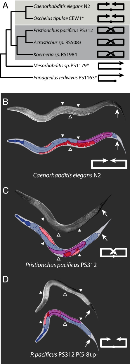
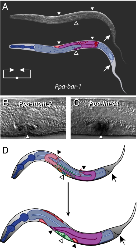
Changes in organ morphology have been essential to the evolution of novel body forms and in permitting organisms to invade new ecological niches. Changes in the arrangement of cells and tissues and in the regulation of morphological movements are fundamental to evolutionary transitions of organ shape and function. However, little is known about the genetic and developmental control of these changes. We use interspecific differences in the migration and extension of the nematode hermaphrodite gonadal arms to study the generation of morphological novelty. We show that the extending Pristionchus pacificus gonadal arms display a ventral migration that is unique to the Diplogastridae in comparison to the Rhabditidae, including Caenorhabditis elegans, and other nematodes. This results in the distal gonad residing along the ventral side of the body in P. pacificus in contrast to lying on the dorsal side of the body as in C. elegans. We show that at the cellular level this morphogenetic movement is regulated by signals from the developing vulva and the sister gonadal arm. We further show that in P. pacificus Wnt signaling is essential for this regulation. We show genetic and molecular evidence that suggest the Wnt ligands Ppa-mom-2 and Ppa-cwn-2 are components of the signaling mechanism. Supporting these findings, the hermaphrodite gonad of Ppa-bar-1 mutant animals mimics the shape of the C. elegans hermaphrodite gonad; the arms fail to extend ventrally. Thus, this genetic analysis of gonad migration provides insight into the mechanisms underlying the generation of morphological novelty and organ shape.
Conflict of interest statement
The authors declare no conflict of interest.
Figures
Similar articles
-
Gonadogenesis in Pristionchus pacificus and organ evolution: development, adult morphology and cell-cell interactions in the hermaphrodite gonad.Dev Biol. 2005 Jan 1;277(1):200-21. doi: 10.1016/j.ydbio.2004.09.021. Dev Biol. 2005. PMID: 15572150
-
Wnt signaling induces vulva development in the nematode Pristionchus pacificus.Curr Biol. 2008 Jan 22;18(2):142-6. doi: 10.1016/j.cub.2007.12.048. Curr Biol. 2008. PMID: 18207741
-
Distinct functions of three Wnt proteins control mirror-symmetric organogenesis in the C. elegans gonad.Elife. 2024 Nov 1;13:e103035. doi: 10.7554/eLife.103035. Elife. 2024. PMID: 39485276 Free PMC article.
-
Nematode model systems in evolution and development.Wiley Interdiscip Rev Dev Biol. 2012 May-Jun;1(3):389-400. doi: 10.1002/wdev.33. Epub 2012 Jan 27. Wiley Interdiscip Rev Dev Biol. 2012. PMID: 23801489 Review.
-
Gonad morphogenesis and distal tip cell migration in the Caenorhabditis elegans hermaphrodite.Wiley Interdiscip Rev Dev Biol. 2012 Jul-Aug;1(4):519-31. doi: 10.1002/wdev.45. Wiley Interdiscip Rev Dev Biol. 2012. PMID: 23559979 Free PMC article. Review.
Cited by
-
Toward Universal Forward Genetics: Using a Draft Genome Sequence of the Nematode Oscheius tipulae To Identify Mutations Affecting Vulva Development.Genetics. 2017 Aug;206(4):1747-1761. doi: 10.1534/genetics.117.203521. Epub 2017 Jun 19. Genetics. 2017. PMID: 28630114 Free PMC article.
-
Genome-wide analysis of trans-splicing in the nematode Pristionchus pacificus unravels conserved gene functions for germline and dauer development in divergent operons.RNA. 2014 Sep;20(9):1386-97. doi: 10.1261/rna.041954.113. Epub 2014 Jul 11. RNA. 2014. PMID: 25015138 Free PMC article.
-
From "the Worm" to "the Worms" and Back Again: The Evolutionary Developmental Biology of Nematodes.Genetics. 2018 Oct;210(2):397-433. doi: 10.1534/genetics.118.300243. Genetics. 2018. PMID: 30287515 Free PMC article. Review.
-
Molecular phylogeny of beetle associated diplogastrid nematodes suggests host switching rather than nematode-beetle coevolution.BMC Evol Biol. 2009 Aug 24;9:212. doi: 10.1186/1471-2148-9-212. BMC Evol Biol. 2009. PMID: 19703296 Free PMC article.
-
β-catenin-dependent Wnt signaling in C. elegans: teaching an old dog a new trick.Cold Spring Harb Perspect Biol. 2012 Aug 1;4(8):a007948. doi: 10.1101/cshperspect.a007948. Cold Spring Harb Perspect Biol. 2012. PMID: 22745286 Free PMC article. Review.
References
-
- Averof M, Cohen SM. Evolutionary origin of insect wings from ancestral gills. Nature. 1997;385:627–630. - PubMed
-
- Damen WG, Saridaki T, Averof M. Diverse adaptations of an ancestral gill: A common evolutionary origin for wings, breathing organs, and spinnerets. Curr Biol. 2002;12:1711–1716. - PubMed
-
- Bishopric NH. Evolution of the heart from bacteria to man. Ann NY Acad Sci. 2005;1047:13–29. - PubMed
-
- Chitwood BG, Chitwood MB. Introduction to Nematology. Baltimore: University Park; 1974.
Publication types
MeSH terms
Substances
LinkOut - more resources
Full Text Sources
