Comparative Study
. 2008 Aug 5;105(31):11020-5.
doi: 10.1073/pnas.0800886105. Epub 2008 Jul 29.

A pH-stabilizing role of voltage-gated proton channels in IgE-mediated activation of human basophils

Affiliations
Comparative Study

A pH-stabilizing role of voltage-gated proton channels in IgE-mediated activation of human basophils

Boris Musset et al. Proc Natl Acad Sci U S A. .

Abstract

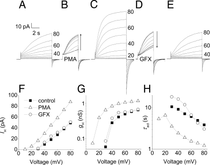
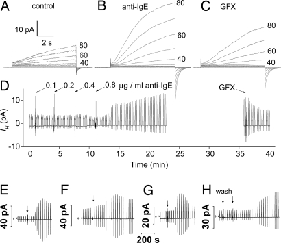
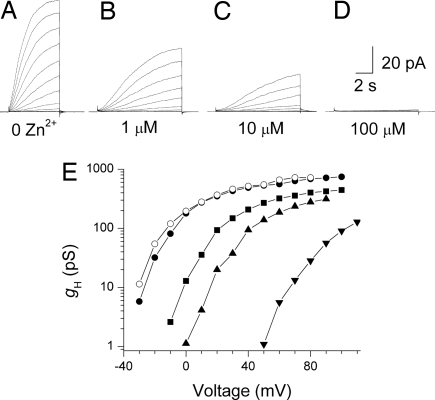
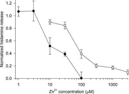
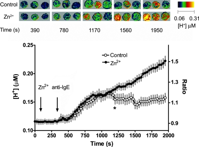
Eosinophils and other phagocytes use NADPH oxidase to kill bacteria. Proton channels in human eosinophils and neutrophils are thought to sustain NADPH oxidase activity, and their opening is greatly enhanced by a variety of NADPH oxidase activators, including phorbol myristate acetate (PMA). In nonphagocytic cells that lack NADPH oxidase, no clear effect of PMA on proton channels has been reported. The basophil is a granulocyte that is developmentally closely related to the eosinophil but nevertheless does not express NADPH oxidase. Thus, one might expect that stimulating basophils with PMA would not affect proton currents. However, stimulation of human basophils in perforated-patch configuration with PMA, N-formyl-methionyl-leucyl-phenylalanine, or anti-IgE greatly enhanced proton currents, the latter suggesting involvement of proton channels during activation of basophils by allergens through their highly expressed IgE receptor (Fc epsilonRI). The anti-IgE-stimulated response occurred in a fraction of cells that varied among donors and was less profound than that to PMA. PKC inhibition reversed the activation of proton channels, and the proton channel response to anti-IgE or PMA persisted in Ca(2+)-free solutions. Zn(2+) at concentrations that inhibit proton current inhibited histamine release elicited by PMA or anti-IgE. Studied with confocal microscopy by using SNARF-AM and the shifted excitation and emission ratioing of fluorescence approach, anti-IgE produced acidification that was exacerbated in the presence of 100 microM Zn(2+). Evidently, proton channels are active in basophils during IgE-mediated responses and prevent excessive acidification, which may account for their role in histamine release.


Conflict of interest statement

The authors declare no conflict of interest.

Figures

Fig. 1.
Fig. 1.
Proton currents in human basophils respond vigorously to PMA and GFX. (A, C, and E) Families of currents in a basophil before stimulation (A), after exposure to 60 nM PMA for 4 min (C), and after addition of 2.5 μM GFX (E). In each, the membrane was held at −20 mV, and 8-s pulses were applied in 10-mV increments every 30 s up to +80 mV. (B) Superimposed currents during the transition to PMA, with 4-s pulses to +60 mV applied every 15 s. (D) Test currents after addition of GFX. (F) I–V relationships from the families in A, C, and E. (G) gHV relationships from the families in A, C, and E were calculated by using Vrev measured in each condition and the steady-state IH obtained by extrapolating exponential fits. (H) Voltage dependence of the activation time constant (τact) obtained by fitting the currents in A, C, and E to a rising exponential (after a delay). Calibration bars apply to A–E. The bath included Ca2+.
Fig. 2.
Fig. 2.
Proton currents in human basophils are enhanced by anti-IgE stimulation in a GFX-sensitive manner. (A–C) Currents in response to the same applied family of pulses before stimulation (A), after stimulation by 0.8 μg/ml anti-IgE (B), and in the presence of 3 μM GFX (C). (D) The time course of these responses. Anti-IgE was added to the bath incrementally, and then the bath stirred. The bath was Ca2+ free. (E–H) Time course of anti-IgE responses in four other basophils. Horizontal arrows indicate zero current. In each, test pulses to +40 mV (E–G) or +60 mV (H) were applied every 30 s, and at the arrow, 0.6 μg/ml anti-IgE was applied to the bath followed by stirring, except for G, where 6.6 μg/ml was applied. In H, “wash” indicates a bath exchange with the same solution as a control; in this experiment anti-IgE was applied by complete bath exchange. The bath included 2 mM Ca2+, except H, which was Ca2+-free.
Fig. 3.
Fig. 3.
Effects of Zn2+ on proton currents in a human basophil at pHo 7.4 (Ringer's solution without EGTA) and pHi 5.5. (A–D) Families of currents in response to depolarizing pulses applied from a holding potential of −60 mV to +60 mV in 10-mV increments. The Zn2+ concentration was 0 (A), 1 μM (B), 10 μM (C), and 100 μM (D). (E) The gHV relationships from this experiment, calculated by using the current at the end of 6-s pulses (control and wash) or 10-s pulses (all Zn2+ concentrations). Symbols in the order of the experiment are ● (control), ■ (1 μM Zn2+), ▴ (10 μM Zn2+), ▾ (100 μM Zn2+), and ○ (after washout). Because Zn2+ slows activation, the current amplitude at the end of the pulse increasingly underestimates the steady-state value at higher [Zn2+]. Pulses to larger voltages are included in E.
Fig. 4.
Fig. 4.
Inhibition of histamine release by Zn2+. Histamine release induced by 0.3 μg/ml anti-IgE (●) or 60 nM PMA (□) in the presence of various concentrations of ZnCl2, normalized to release in the absence of ZnCl2. Each value is the mean ± SEM of measurements in three to five experiments using blood from different donors. Spontaneous release was 10.0 ± 0.9% (mean ± SEM, n = 5) of total histamine content (range 7.7–12.9%), the PMA stimulated release was 65 ± 7% (42–82%, n = 5 except n = 3 at 10 μM Zn2+), and the anti-IgE stimulated release was 24 ± 6% (13–34%, n = 3). The values for the two stimuli at 30 μM and 100 μM differ significantly (P < 0.001 and P < 0.05, respectively, by Student's t test).
Fig. 5.
Fig. 5.
Average [H+]i in basophils (99% pure) stimulated with 1 μg/ml anti-IgE in the absence (○) or presence of 100 μM Zn2+ (●) at ≈30°C and imaged by using confocal microscopy and SEER. The mean ± SEM of 25 control cells and 46 cells in Zn2+ is shown, with all data pairs after the star significantly different by Student's t test (P < 0.05). Pseudocolor images indicating [H+]i in trios of basophils from this experiment taken after stimulation with anti-IgE at the indicated time points are shown in two rows; the top row is control, the lower in the presence of Zn2+.

Similar articles

Cited by

References

    1. Cherny VV, Thomas LL, DeCoursey TE. Voltage-gated proton currents in human basophils. Biol Membr. 2001;18:458–465.
    1. DeCoursey TE. Voltage-gated proton channels and other proton transfer pathways. Physiol Rev. 2003;83:475–579. - PubMed
    1. Henderson LM, Chappell JB, Jones OTG. The superoxide-generating NADPH oxidase of human neutrophils is electrogenic and associated with an H+ channel. Biochem J. 1987;246:325–329. - PMC - PubMed
    1. DeCoursey TE, Morgan D, Cherny VV. The voltage dependence of NADPH oxidase reveals why phagocytes need proton channels. Nature. 2003;422:531–534. - PubMed
    1. Bánfi B, et al. A novel H+ conductance in eosinophils: Unique characteristics and absence in chronic granulomatous disease. J Exp Med. 1999;190:183–194. - PMC - PubMed

Publication types