. 2008 Aug 6;100(15):1092-103.
doi: 10.1093/jnci/djn216. Epub 2008 Jul 29.

Effect of lapatinib on the outgrowth of metastatic breast cancer cells to the brain

Affiliations

Effect of lapatinib on the outgrowth of metastatic breast cancer cells to the brain

Brunilde Gril et al. J Natl Cancer Inst. .

Abstract

Background: The brain is increasingly being recognized as a sanctuary site for metastatic tumor cells in women with HER2-overexpressing breast cancer who receive trastuzumab therapy. There are no approved or widely accepted treatments for brain metastases other than steroids, cranial radiotherapy, and surgical resection. We examined the efficacy of lapatinib, an inhibitor of the epidermal growth factor receptor (EGFR) and HER2 kinases, for preventing the outgrowth of breast cancer cells in the brain in a mouse xenograft model of brain metastasis.

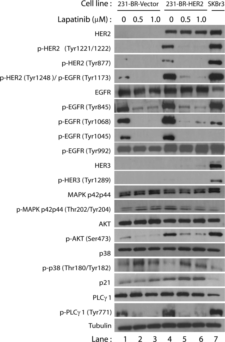
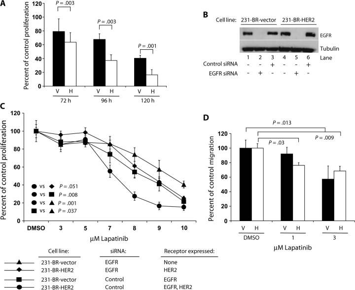
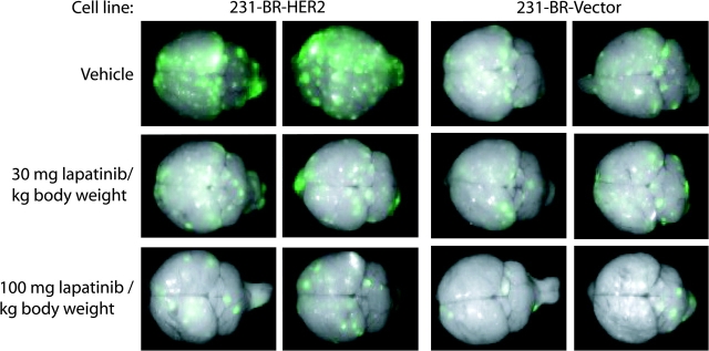
Methods: EGFR-overexpressing MDA-MB-231-BR (231-BR) brain-seeking breast cancer cells were transfected with an expression vector that contained or lacked the HER2 cDNA and used to examine the effect of lapatinib on the activation (ie, phosphorylation) of cell signaling proteins by immunoblotting, on cell growth by the tetrazolium salt 3-(4,5-dimethylthiazol-2-yl)-2,5-diphenyl tetrazolium bromide assay, and on cell migration using a Boyden chamber assay. The outgrowth of large (ie, >50 microm(2)) and micrometastases was counted in brain sections from nude mice that had been injected into the left cardiac ventricle with 231-BR cells and, beginning 5 days later, treated by oral gavage with lapatinib or vehicle (n = 22-26 mice per treatment group). All statistical tests were two-sided.

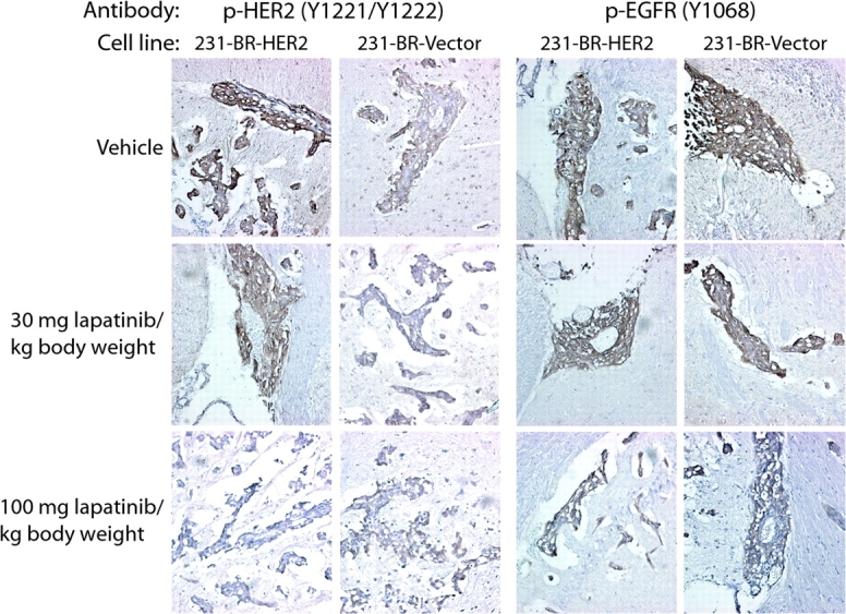
Results: In vitro, lapatinib inhibited the phosphorylation of EGFR, HER2, and downstream signaling proteins; cell proliferation; and migration in 231-BR cells (both with and without HER2). Among mice injected with 231-BR-vector cells, those treated with 100 mg lapatinib/kg body weight had 54% fewer large metastases 24 days after starting treatment than those treated with vehicle (mean number of large metastases per brain section: 1.56 vs 3.36, difference = 1.80, 95% confidence interval [CI] = 0.92 to 2.68, P < .001), whereas treatment with 30 mg lapatinib/kg body weight had no effect. Among mice injected with 231-BR-HER2 cells, those treated with either dose of lapatinib had 50%-53% fewer large metastases than those treated with vehicle (mean number of large metastases per brain section, 30 mg/kg vs vehicle: 3.21 vs 6.83, difference = 3.62, 95% CI = 2.30 to 4.94, P < .001; 100 mg/kg vs vehicle: 3.44 vs 6.83, difference = 3.39, 95% CI = 2.08 to 4.70, P < .001). Immunohistochemical analysis revealed reduced phosphorylation of HER2 in 231-BR-HER2 cell-derived brain metastases from mice treated with the higher dose of lapatinib compared with 231-BR-HER2 cell-derived brain metastases from vehicle-treated mice (P < .001).

Conclusions: Lapatinib is the first HER2-directed drug to be validated in a preclinical model for activity against brain metastases of breast cancer.


Figures

Figure 1
Figure 1
Effect of lapatinib on expression of proteins involved in HER2 and epidermal growth factor receptor (EGFR) signaling pathways in 231-BR cells. 231-BR-vector control (lanes 1–3) or 231-BR-HER2 (lanes 4–6) cells were serum starved overnight and subsequently treated with either 0.5 or 1.0 μM lapatinib for 24 h. Lapatinib was diluted in dimethyl sulfoxide, which served as the vehicle control (lanes 1 and 4). After lapatinib or vehicle treatment, cells were stimulated with 100 ng/mL epidermal growth factor for 10 min and total cell lysates were then prepared for immunoblot analysis. SKBr3 cells (lane 7), which endogenously overexpress HER2, were used as a positive control for antibody detection of HER family members. Proteins analyzed were HER2, EGFR, HER3, mitogen activated protein kinase (MAPK p42/p44), AKT, p38, p21, PLCγ1, tubulin. The prefix “p-” refers to phosphorylation of the residues in parentheses. The data shown are representative of results obtained in at least three separate experiments.
Figure 2
Figure 2
Effects of lapatinib on 231-BR cell proliferation and migration. A) Time course of lapatinib (8 μM) inhibition of the proliferation of 231-BR-vector (V)– and 231-BR-HER2 (H)–overexpressing cell lines. The mean percentage proliferation versus diluent (dimethyl sulfoxide, DMSO) control treatment for each cell line and time point is shown, and error bars represent 95% confidence intervals. B) and C) Dependence of the antiproliferative activity of lapatinib on the number of target receptors expressed. 231-BR-vector cells or 231-BR-HER2 cells were incubated with small interfering RNA (siRNA) targeting EGFR or a nontargeting control (a sequence not homologous to any human, mouse, or rat gene) siRNA. After 24 h of siRNA treatment, cells were cultured in the presence of various doses of lapatinib or diluent (DMSO) for 96 h. Half of the diluent-treated cells were lysed for immunoblot analysis of epidermal growth factor receptor expression (B), and the remaining cells were assayed for cell viability using the 3-(4,5-dimethylthiazol-2-yl)-2,5-diphenyl tetrazolium bromide assay (C). The P values represent data averaged over the 7- to 10-μM doses of lapatinib. D) In vitro cell migration assay. 231-BR-vector (V) or 231-BR-HER2 cells (H) were pretreated with 1 or 3 μM lapatinib or diluent (DMSO). Cell migration in response to 1.0% fetal bovine serum was examined using a Boyden chamber assay. The graph represents percentage of migration compared with DMSO control treatment. For the 231-BR-vector cell line, no statistically significant difference was observed between DMSO and 1 μM lapatinib treatment (P = .27). All P values are two-sided (analysis of variance).
Figure 3
Figure 3
Lapatinib inhibition of metastatic colonization of mouse brain by 231-BR breast carcinoma cells. 231-BR-HER2 cells or 231-BR-vector cells, both of which were transduced with a retrovirus that expressed enhanced green fluorescent protein (EGFP), were injected into the left cardiac ventricle of BALB/c nude mice. Five days after injection, lapatinib (30 or 100 mg/kg body weight) or vehicle (0.5% hydroxypropylmethylcellulose with 0.1% Tween 80 in water) was administered by twice-daily oral gavage for 24 days (n = 22–26 mice per treatment group). Brains dissected at necropsy were imaged using a Maestro 420 Spectral Imaging System to detect the presence of EGFP expressing metastases derived from the injected 231-BR cells (metastatic foci on a green to white [greater intensity] fluorescent intensity spectrum). Representative dorsal whole-brain images from two mice in each treatment group are shown.
Figure 4
Figure 4
Immunohistochemical evaluation of HER2 and epidermal growth factor receptor (EGFR) activation in vivo in response to lapatinib treatment. Frozen sections (5 µm thick) of brains from mice injected with 231-BR-vector or 231-BR-HER2 cells and treated with lapatinib (30 or 100 mg/kg body weight) or vehicle (n = 5 mice per group) were stained with antibodies specific for phosphorylated HER2 (p-HER2; tyrosines 1221 and 1222) or phosphorylated EGFR (p-EGFR; tyrosine 1068). The staining of all large metastases (ie, >50 μm2) and 125 micrometastases per treatment group was scored on a 0–3+ intensity scale by two investigators who were blinded to the treatment group assignment. Representative images of large metastases for each group are shown (×200 magnification). The presence of p-HER2 or p-EGFR antigen is indicated by brown staining; nuclei were counterstained purple with hematoxylin.

References

    1. Weil R, Palmieri D, Bronder J, Stark A, Steeg P. Breast cancer metastasis to the central nervous system. Am J Pathol. 2005;167(4):913–920. - PMC - PubMed
    1. Lin N, Bellon J, Winer E. CNS metastases in breast cancer. J Clin Oncol. 2004;22(17):3608–3617. - PubMed
    1. Slotman B, Faive-Finn C, Kramer G, et al. Prophylactic cranial irradiation in extensive small-cell lung cancer. N Engl J Med. 2007;357(7):664–672. - PubMed
    1. Sneed PK, Lamborn KR, Forstner JM, et al. Radiosurgery for brain metastases: is whole brain radiotherapy necessary? Int J Radiat Oncol Biol Phys. 1999;43(3):549–558. - PubMed
    1. Patchell RA, Regine WF. The rationale for adjuvant whole brain radiation therapy with radiosurgery in the treatment of single brain metastases. Technol Cancer Res Treat. 2003;2(2):111–115. - PubMed

Publication types

MeSH terms