. 2008 Jul 30;3(7):e2831.
doi: 10.1371/journal.pone.0002831.

The N-terminal domain of the Drosophila retinoblastoma protein Rbf1 interacts with ORC and associates with chromatin in an E2F independent manner

Affiliations

The N-terminal domain of the Drosophila retinoblastoma protein Rbf1 interacts with ORC and associates with chromatin in an E2F independent manner

Joseph Ahlander et al. PLoS One. .

Abstract

Background: The retinoblastoma (Rb) tumor suppressor protein can function as a DNA replication inhibitor as well as a transcription factor. Regulation of DNA replication may occur through interaction of Rb with the origin recognition complex (ORC).

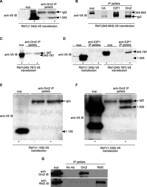
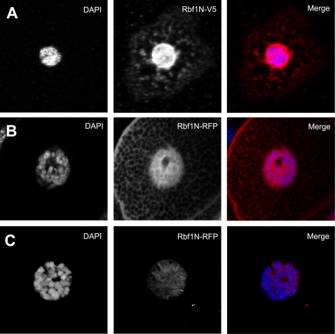
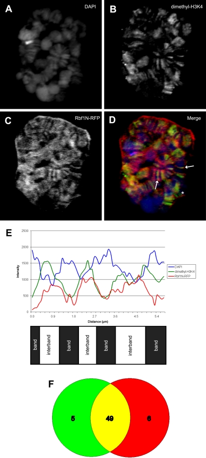
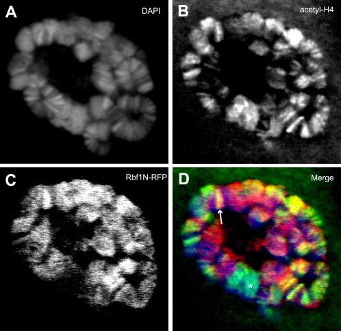
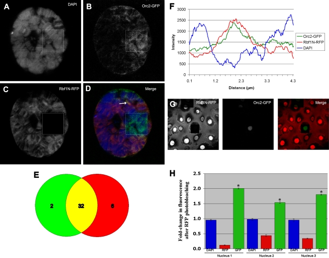
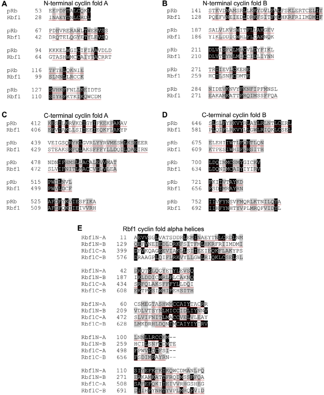
Principal findings: We characterized the interaction of Drosophila Rb, Rbf1, with ORC. Using expression of proteins in Drosophila S2 cells, we found that an N-terminal Rbf1 fragment (amino acids 1-345) is sufficient for Rbf1 association with ORC but does not bind to dE2F1. We also found that the C-terminal half of Rbf1 (amino acids 345-845) interacts with ORC. We observed that the amino-terminal domain of Rbf1 localizes to chromatin in vivo and associates with chromosomal regions implicated in replication initiation, including colocalization with Orc2 and acetylated histone H4.

Conclusions/significance: Our results suggest that Rbf1 can associate with ORC and chromatin through domains independent of the E2F binding site. We infer that Rbf1 may play a role in regulating replication directly through its association with ORC and/or chromatin factors other than E2F. Our data suggest an important role for retinoblastoma family proteins in cell proliferation and tumor suppression through interaction with the replication initiation machinery.


Conflict of interest statement

Competing Interests: The authors have declared that no competing interests exist.

Figures

Figure 1
Figure 1. ORC interacts with Rbf1 N-terminal and C-terminal fragments in an E2F independent manner.
S2 cells were transfected with metallothionein promoter (pMT) regulated Rbf1 deletion constructs with a C-terminal Simian Virus 5 (V5) epitope-tag and cell extracts from uninduced (−) and copper sulfate induced (+) cultures were subjected to immunoprecipitation (IP) and anti-V5 immunoblotting (IB). (A) Rbf1(1–345)-V5 transfected cell extracts were immunoprecipitated with anti-Orc2. Note that extracts from induced cells show a Rbf1(1–345)-V5 fragment (arrowhead) in the IP pellet while uninduced cell extracts treated identically with anti-Orc2 serum failed to IP an anti-V5 reacting band of comparable size. (B) Rbf1(345–845)-V5 transfected cell extracts were induced and anti-HA (negative control), anti-dE2F1 and anti-Orc2 serum were used in IP reactions. Western IB was probed with anti-V5 which detects the Rbf1(345–845)-V5 protein fragment (arrowhead) that migrates just above the IgG heavy chain (arrow). (C) Extracts from Rbf1(345–797)-V5 cells uninduced (−) and induced (+) cultures were subjected to anti-Orc2 IP and western IB probed with anti-V5. (D) Extracts from Rbf1(1–345)-V5 or Rbf1(345–797)-V5 cells uninduced (−) and induced (+) were subjected to anti-dE2F1 IP. Extracts from (E) Rbf1(1–150)-V5 and (F) Rbf1(1–330)-V5 cells that were uninduced (−) and induced (+) were subjected to anti-Orc2 IP and anti-V5 western IB. In each case 5–10% of the IP supernatant (sup.) and all of the IP pellets were loaded. In all panels (except D) the IgG heavy chain protein is noted by an arrow and Rbf1-V5 deletion fragments are denoted by an arrowhead. (G) Rbf2 does not interact with ORC. Ovarian extracts were immunoprecipitated (IP pellets) with no antibody (No Ab.), anti-Orc2 or anti-Rbf2. Entire IP pellets and 10% of supernatant were loaded. Immunoblot (IB) was first probed with anti-Orc2, stripped and then reprobed with anti-Rbf2.
Figure 2
Figure 2. The Rbf1 amino-terminal domain, Rbf1N, is sufficient for nuclear localization and chromatin association.
(A) S2 cells were transfected with a copper inducible construct containing Rbf1N (Rbf1 amino acids 1–345) tagged with a V5 epitope. Immunofluorescence using V5 antibodies shows Rbf1N is mostly nuclear with small amounts cytoplasmic localization. To observe the localization of Rbf1N in vivo, transgenic flies containing UAS>Rbf1N-RFP were crossed to flies bearing a GAL4 transgene that expressed specifically in salivary glands. (B) Rbf1N-RFP localizes to the nucleus in salivary gland cells. RFP fluorescence is brightly seen throughout the nucleus and cytoplasm, and it appears to also associate with cytoplasmic structures and the plasma membrane. (C) To remove unbound Rbf1N-RFP, salivary glands were incubated in chromatin wash buffer, revealing that Rbf1N-RFP associates with chromatin and localizes in a striped pattern along polytene chromosomes.
Figure 3
Figure 3. Rbf1N colocalizes with modified histones at interband regions of salivary gland polytene chromosomes.
(A) Salivary glands expressing Rbf1N-RFP were chromatin washed and counterstained with antibodies specific for histone H3 dimetylated on lysine 4, a modified histone that marks interband DNA and is an indicator of active transcription. Rbf1N-RFP (C) and dimethyl-H3K4 (B) colocalize at interbands (D and E), whereas DAPI stains the bands of the polytene chromosomes (A and E). Arrows indicate interbands demonstrating colocalization, and the asterisk denotes a site where colocalization does not occur. The merged image (D) reveals extensive colocalization of Rbf1N-RFP and dimethyl-H3K4, as well as some areas of non-overlap. (E) A graph of fluorescent intensity along several chromosome bands shows the banding pattern of DAPI versus the alternating interband pattern of Rbf1N-RFP and dimethyl-H3K4. (F) A Venn diagram illustrates that Rbf1N-RFP colocalizes extensively with the modified histone dimethyl-H3K4 in randomly chosen bands.
Figure 4
Figure 4. Rbf1N colocalizes with acetylated histone H4 at interband regions of salivary gland polytene chromosomes.
Salivary glands expressing Rbf1N-RFP were chromatin washed and counterstained with antibodies specific for acetylated histone H4, a marker of active transcription and active origins of replication. Acetyl-H4 colocalizes with Rbf1N-RFP at interbands. (A) DAPI staining marks chromosomal bands. Acetyl-H4 (B) and Rbf1N-RFP (C) colocalize at many chromosomal locations (D). The merged image (D) reveals extensive colocalization of Rbf1N-RFP and acetyl-H4, as well as some areas of non-overlap. The arrow indicates an interband representing colocalization, and the asterisk denotes a site where colocalization does not occur.
Figure 5
Figure 5. Rbf1N physically interacts with ORC in vivo.
Salivary glands from transgenic larvae expressing both Rbf1N-RFP and Orc2-GFP were chromatin washed and fixed for fluorescence microscopy. Rbf1N-RFP (C) and Orc2-GFP (B) colocalize on polytene chromosomes (D through F). DAPI stains the bands of the polytene chromosomes (A). Photobleaching of Rbf1N-RFP, indicated by the boxed area, results in an increased GFP signal, which is a consequence of fluorescence resonance energy transfer (FRET) by the red and green fluorescent proteins, mCherry and EGFP. FRET reveals that Rbf1N-RFP and Orc2-GFP are in very close physical proximity. (E) A Venn diagram illustrates that Orc2-GFP colocalizes extensively with Rbf1N-RFP fluorescence in randomly chosen bands. (F) A graph of fluorescent intensity along the region indicated by an arrow (D) shows that Rbf1N-RFP and Orc2-GFP colocalize within an interband region. (G) Photobleaching of RbfN-RFP results in an increased GFP signal in salivary gland nuclei. (H) Fold-change after Rbf1-N-RFP photobleaching is shown as the ratio of bleached/non-bleached signal in each of three different nuclei. Blue bars show DAPI signal, red is RFP signal and green show fold-change in Orc2-GFP signal. A two-tailed T-test indicates that GFP fluorescence increase is highly statistically significant p<0.0001 in each of the three nuclei. RFP photobleaching increases GFP fluorescence by 1.5–2-fold. These three nuclei (see Figure S1) are representative of larger populations.
Figure 6
Figure 6. Alignment of Cyclin fold helices within the Rbf1 sequence.
The retinoblastoma proteins in humans and flies share a domain structure containing four cyclin folds, with each fold consisting of five alpha helices. The N-terminal (A and B) and C-terminal (C and D) domains of Rbf1 each have a cyclin fold A and B, resulting in four total cyclin folds that share extensive sequence conservation with pRb. It is likely that the retinoblastoma family of proteins emerged from two successive tandem duplication events from an ancient cyclin-like ancestor that gave rise to many cell cycle regulators. This finding seems to indicate that the retinoblastoma N and C-terminal domains are intrahomologues. The tandem domain architecture of Rb family proteins may explain our finding that ORC interacts with multiple Rbf1 domains, and suggests that Rbf1 may be an adaptor molecule that is able to switch between several orientations with ORC to accommodate different combinations of binding partners depending on different cellular contexts. (E) All five helices from the four Rbf1 cyclin folds were compared together. Amino acids conserved in two or more helices were shaded accordingly, revealing a collective conservation of amino acid sequence between the cyclin folds. Black shading with white letters indicates identical amino acids. Grey shading indicates amino acid similarity. Helices are underlined in red.
Figure 7
Figure 7. Models of Rbf1 adaptor functions.
(A) Rbf1 associates with ORC and may inhibit recruitment other replication initiation factors. Due to its association with ORC, Rbf1 might inhibit the activity of the replication initiation complex. Phosphorylation of the C-terminal domain of Rbf1 by Cyclin-CDK complexes releases binding partners, such as E2F, and may constitute part of a reversible switch to regulate origins of replication. This switchable regulation may come in part through changes in recruitment of associated chromatin modifying enzymes and tethering of phosphorylated Rbf1 by the Myb-MuvB complex may allow Rbf1 to ping-pong from one complex to another in a localized manner. (B) We speculate that because Rbf1 may be able to associate with chromatin bound ORC and through multiple domains it can be tethered in more than one orientation, thereby presenting and/or occluding docking sites for other Rbf1-associated chromatin factors (e.g. histone deacetylases, histone methyltransferase, etc.). For example, this may allow Rbf1 to function as an “adaptor” molecule at any one ORC site where its specific orientation dictates which factors (depicted as “X” and “Y”) may or may not be present at any given time. This model predicts that a single genomic site may have constitutive ORC/Rbf1 localization while re-orientation of the Rbf1 molecule can mediate the recruitment of different suites of chromatin modifying enzymes. This model and that described above (A) are not mutually exclusive.

References

    1. Machida YJ, Hamlin JL, Dutta A. Right place, right time, and only once: Replication initiation in metazoans. Cell. 2005;123(1):13–24. - PubMed
    1. Tanny RE, MacAlpine DM, Blitzblau HG, Bell SP. Genome-wide analysis of re-replication reveals inhibitory controls that target multiple stages of replication initiation. Mol Biol Cell. 2006;17(5):2415–2423. - PMC - PubMed
    1. Diffley JF. Once and only once upon a time: Specifying and regulating origins of DNA replication in eukaryotic cells. Genes Dev. 1996;10(22):2819–2830. - PubMed
    1. Nowak MA, Komarova NL, Sengupta A, Jallepalli PV, Shih I, et al. The role of chromosomal instability in tumor initiation. Proc Natl Acad Sci U S A. 2002;99(25):16226–16231. - PMC - PubMed
    1. Knudsen ES, Sexton CR, Mayhew CN. Role of the retinoblastoma tumor suppressor in the maintenance of genome integrity. Curr Mol Med. 2006;6(7):749–757. - PubMed

Publication types

LinkOut - more resources