. 2009 Jan;19(1):121-31.
doi: 10.1007/s00330-008-1111-x. Epub 2008 Jul 30.

Magnetic resonance imaging for monitoring the effects of thalidomide on experimental human breast cancers

Affiliations

Magnetic resonance imaging for monitoring the effects of thalidomide on experimental human breast cancers

Clemens C Cyran et al. Eur Radiol. 2009 Jan.

Abstract

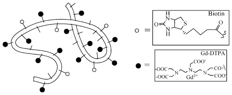
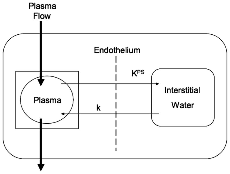
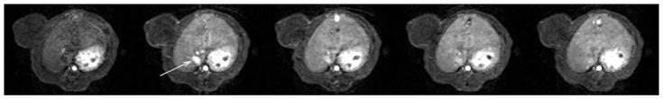
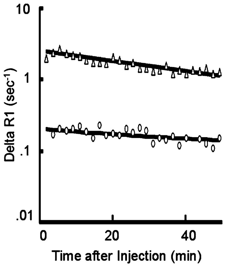
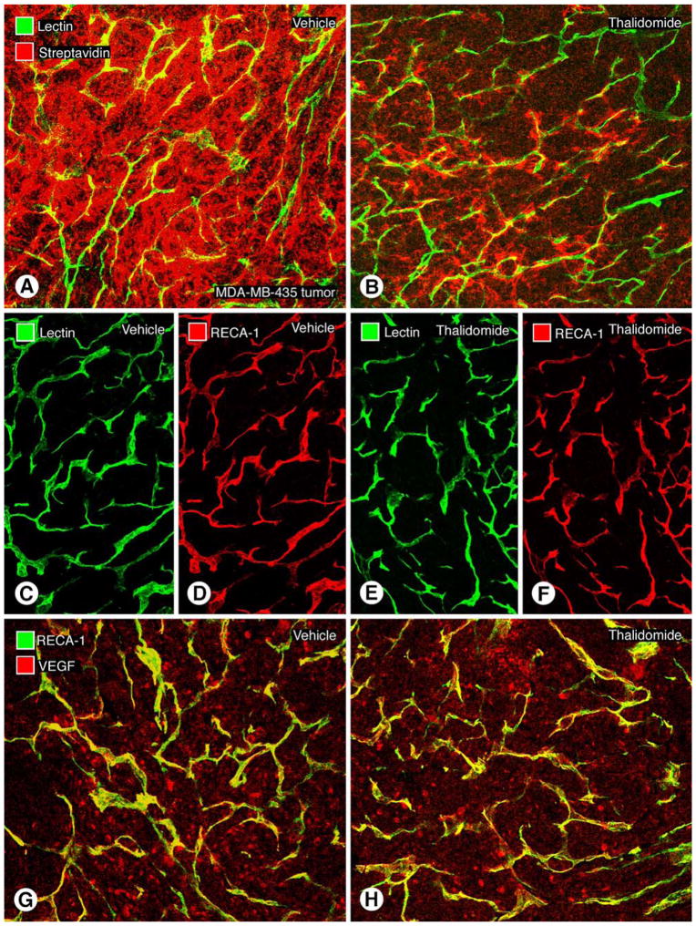
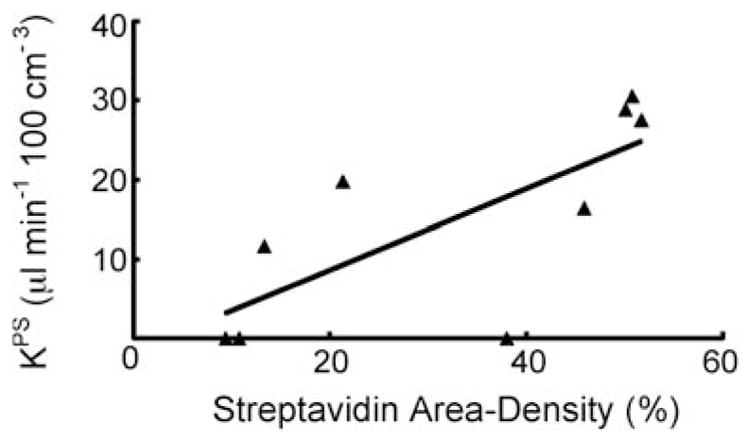
Thalidomide, which inhibits angiogenesis in certain tumor types, reduced extravasation of a macromolecular contrast medium (MMCM) in a human breast cancer model as assayed by MMCM-enhanced dynamic magnetic resonance imaging (MRI) and fluorescence microscopy in the same tumors. After a 1-week, three-dose course of thalidomide, the mean MRI-assayed endothelial transfer coefficient, K(PS), decreased significantly (p < 0.05) from 19.4 +/- 9.1 to 6.3 +/- 9.1 microl/min.100 cm(3). Correspondingly, microscopic measurements of extravasated MMCM, expressed as fractional area of streptavidin staining, were significantly (p < 0.05) lower in thalidomide-treated tumors (18.6 +/- 11.9%) than in control saline-treated tumors (50.2 +/- 2.3%). On a tumor-by-tumor basis, post-treatment K(PS) values correlated significantly (r(2) = 0.55, p < 0.05) with microscopic measures of MMCM extravasation. However, no significant differences were observed between saline- and thalidomide-treated tumors with respect to rate of growth, vascular richness, or amount of VEGF-containing cells. Because of its sensitivity to the detection of changes in vascular leakage in tumors, this MMCM-enhanced MRI assay could prove useful for monitoring the effects of thalidomide on an individual patient basis. The significant correlation between MRI and fluorescence microscopic measures of MMCM extravasation supports the utility of the non-invasive MRI approach for assessing the action of thalidomide on tumor blood vessels.


Figures

Fig. 1
Fig. 1
Schematic structure of albumin-(Gd-DTPA)27-(biotin)11. The belt-like random coil represents the human albumin core element with amino-containing side chains of the lysine residues (small sticks) to which (Gd-DTPA) (black spheres) and biotin (white spheres) are covalently attached
Fig. 2
Fig. 2
Kinetic model. A simple two-compartment tissue model describing the kinetics of contrast media transport from the plasma space into the interstitial fluid. The endothelial transfer coefficient KPS [μl/(min·100 cm3)] denotes the clearance of contrast medium from plasma to interstitial water. Denoting the fractional rate of reflux from interstitial fluid back to plasma, the rate constant k (min−1) was not resolvable in the time course of these experiments (1 h) and is therefore set to zero. The box around the plasma compartment denotes a forcing function, representing the monoexponential disappearance of MMCM from the blood. The kinetics of both compartments, taken together, reflect the dynamic response of the entire tissue/tumor to contrast medium enhancement following bolus intravenous injection
Fig. 3
Fig. 3
Representative T1-weighted spoiled gradient refocused (SPGR) images, precontrast and at 3 min, 15 min, 30 min and 60 min post injection of albumin-(Gd-DTPA)27-(biotin)11, which were used for the calculation of the endothelial transfer coefficient (KPS) and the fractional plasma volume (fPV). Note the tumor enhancement, most prominent in the rim. Due to the macromolecular nature of albumin-(Gd-DTPA)11-biotin27,however, the first pass extraction fraction in tumor tissue of our contrast agent is in the order of less than 0.1% and the changes in signal intensity caused thereby are correspondingly small. The blood enhancement seen in the IVC (white arrow) persisted over the 1-h course of data acquisition
Fig. 4
Fig. 4
Representative fit (solid lines) of model to ΔR1 data from blood (Δ) and tumor (O) using MMCM enhancement. The deviation from a parallel relationship toward convergence of the blood and tumor fits on a semi-logarithmic plot indicates a leak of the MMCM from the blood into the interstitial space of this tumor. Had the two lines been parallel, it would have indicated no measurable leak. Due to the macromolecular nature of albumin-(Gd-DTPA)27-biotin11, and the therefore low first-pass extraction fraction in tumor tissue, the convergence of the two lines on the semi-logarithmic plot can be somewhat difficult to appreciate visually, in this case of a discrete leak
Fig. 5
Fig. 5
Representative immunohistochemically stained human breast cancer sections (MDA-MB-435) showing leakage of macromolecular contrast medium (streptavidin-biotin reaction), vascular richness (lectin and RECA-1 antibody), and VEGF in tumor cells. Thalidomide treatment reduces the extravasation of albumin-(Gd-DTPA)27-(biotin)11 without reducing the abundance of tumor blood vessels. a Tumor section from the saline-control group administered albumin-(Gd-DTPA)27-(biotin)11. The strong red signal indicates extravascular 1-h accumulation of biotin-labeled contrast medium surrounding the yellow-green tumor microvessels. b After 7 days of treatment with thalidomide, the density of red-fluorescent, biotin-labeled contrast agent extravasated over 1 h is strongly reduced compared with a, indicating a reduction in leakage and extravascular accumulation of the macromolecules. Confocal microscopic images of tumor vessels in MDA-MB-435 tumors after treatment with saline (c, d) or thalidomide (e, f) for 7 days show no noticeable change in the area density of perfused blood vessels (green lectin-stained) or total blood vessels (red, RECA-1 stained). No difference is observable (cf) between saline-control and thalidomide-treated groups with regard to tumor vascularity. (g, h). Representative MDA-MB-435 tumor sections after immunohistochemical staining for human VEGF after a 7-day, three-injection treatment protocol with saline (g) or thalidomide (h). No difference in amount VEGF immunoreactivity was detected in the two groups. Scale bar 115 μm in cf and 120 μm in a, b
Fig. 6
Fig. 6
Graph showing the significant positive correlation (r=0.74, r2=0.55) for each examined tumor between the MRI-assayed endothelial transfer coefficient KPS [μl/(min·100 cm3)] and the immunohistochemically assessed streptavidin area density (%). Both KPS and streptavidin area density reflect the extent of leaked contrast medium. The solid line denotes the best fit

References

    1. Mellin GW, Katzenstein M. The saga of thalidomide. Neuropathy to embryopathy, with case reports of congenital anomalies. N Engl J Med. 1962;267:1184–1192. - PubMed
    1. D’Amato RJ, Loughnan MS, Flynn E, Folkman J. Thalidomide is an inhibitor of angiogenesis. Proc Natl Acad Sci USA. 1994;91(9):4082–4085. - PMC - PubMed
    1. Bolke E, Gripp S, Peiper M, Budach W, Schwarz A, Orth K, Reinecke P, van de Nes JA. Multifocal epithelioid hemangioendothelioma: case report of a clinical chamaeleon. Eur J Med Res. 2006;11(11):462–466. - PubMed
    1. Klausner JD, Freedman VH, Kaplan G. Thalidomide as an anti-TNF-alpha inhibitor: implications for clinical use. Clin Immunol Immunopathol. 1996;81(3):219–223. - PubMed
    1. Moreira AL, Sampaio EP, Zmuidzinas A, Frindt P, Smith KA, Kaplan G. Thalidomide exerts its inhibitory action on tumor necrosis factor alpha by enhancing mRNA degradation. J Exp Med. 1993;177(6):1675–1680. - PMC - PubMed

Publication types