

MR susceptibility weighted imaging (SWI) complements conventional contrast enhanced T1 weighted MRI in characterizing brain abnormalities of Sturge-Weber Syndrome
- PMID: 18666142
- PMCID: PMC2678730
- DOI: 10.1002/jmri.21435
MR susceptibility weighted imaging (SWI) complements conventional contrast enhanced T1 weighted MRI in characterizing brain abnormalities of Sturge-Weber Syndrome
Abstract
Purpose: To evaluate the efficacy of susceptibility weighted imaging (SWI) in comparison to standard T1 weighted postgadolinium contrast (T1-Gd) MRI in patients with Sturge-Weber Syndrome (SWS).
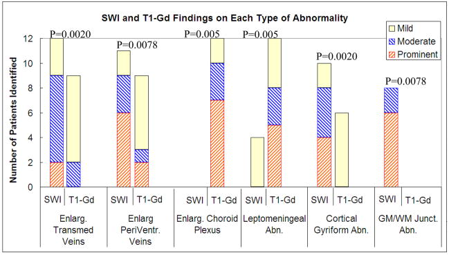
Materials and methods: Twelve children (mean age, 5.6 years) with the diagnosis of SWS and unilateral hemispheric involvement were recruited prospectively and examined with high resolution three dimensional SWI and conventional T1-Gd. Both SWI and T1-Gd images were evaluated using a four-grade scoring system according to six types of imaging findings (enlargement of transmedullary veins, periventricular veins, and choroid plexus, as well as leptomeningeal abnormality, cortical gyriform abnormality, and gray matter/white matter junctional abnormality). The scores of SWI versus T1-Gd images were then compared for each type of abnormality.
Results: SWI was superior to T1-Gd in identifying the enlarged transmedullary veins (P = 0.0020), abnormal periventricular veins (P = 0.0078), cortical gyriform abnormalities (P = 0.0020), and gray matter/white matter junction abnormalities (P = 0.0078). Conversely, T1-Gd was better than SWI in identifying enlarged choroid plexus (P = 0.0050) and leptomeningeal abnormalities (P = 0.0050).
Conclusion: SWI can provide useful and unique information complementary to conventional contrast enhanced T1 weighted MRI for characterizing SWS. Therefore, SWI should be integrated into routine clinical MRI protocols for suspected SWS.
(c) 2008 Wiley-Liss, Inc.
Figures

References
-
- Lee JS, Asano E, Muzik O, Chugani DC, Juhasz C, Pfund Z, Philip S, Behen M, Chugani HT. Sturge-Weber syndrome: correlation between clinical course and FDG PET findings. Neurology. 2001;57:189–95. - PubMed
-
- Fischbein NJ, Barkovich AJ, Wu Y, Berg BO. Sturge-Weber syndrome with no leptomeningeal enhancement on MRI. Neuroradiology. 1998;40:177–180. - PubMed
-
- Reid DE, Maria BL, Drane WE, Quisling RG, Hoang KB. CNS perfusion and metabolism abnormalities in Sturge-Weber syndrome. Journal of Child Neurology. 1997;12(3):218–222. - PubMed
-
- Haacke EM, Xu Y, Cheng YC, Reichenbach JR. Susceptibility weighted imaging (SWI) Magn Res Med. 2004;52:612–8. - PubMed
-
- Reichenbach JR, Venkatesan R, Schillinger DJ, Kido DK, Haacke EM. Small vessels in the human brain: MR venography with deoxyhemoglobin as an intrinsic contrast agent. Radiology. 1997;204:272–279. - PubMed
Publication types
MeSH terms
Substances
Grants and funding
LinkOut - more resources
Full Text Sources
Medical
