

"Melanosis" in the small and large intestine
- PMID: 18666316
- PMCID: PMC2731179
- DOI: 10.3748/wjg.14.4296
"Melanosis" in the small and large intestine
Abstract
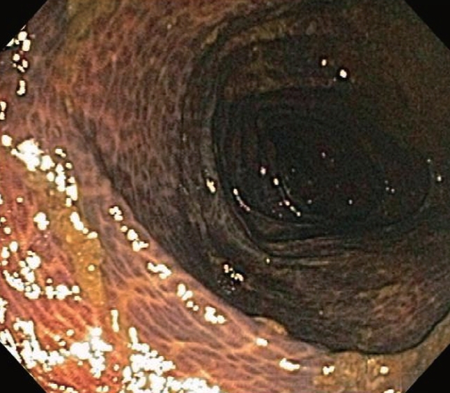
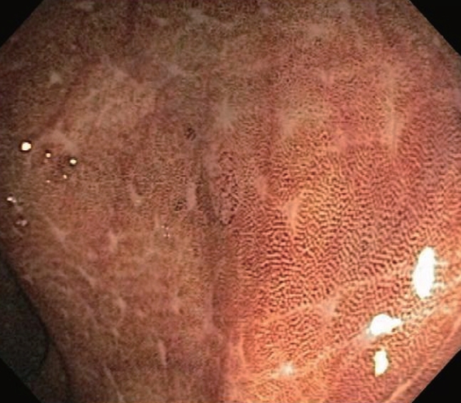
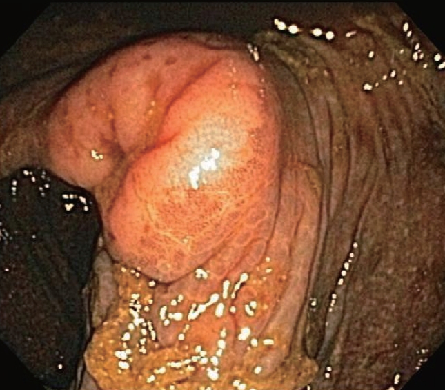
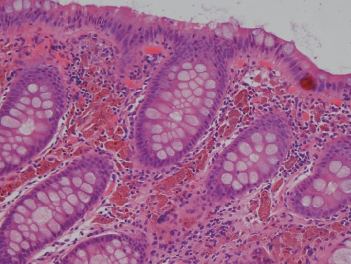
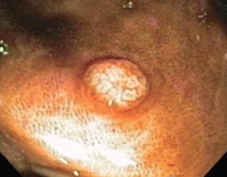
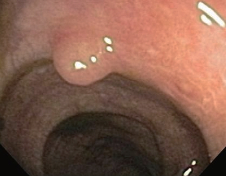
Deposition of pigment in the intestinal mucosa is commonly observed by the endoscopist, especially within the colon, and particularly during investigations for constipation. Pigment may also be detected in the small intestine. Although labeled as melanosis, electron microscopy and X-ray analytical methods have provided evidence that this pigment is not melanin at all, but lipofuscin. Often, herbal remedies or anthracene containing laxatives are often historically implicated, and experimental studies in both humans and animal models have also confirmed the intimate relationship with these pharmacological or pseudo-pharmacological remedies. The appearance of melanosis coli during colonoscopy is largely due to pigment granule deposition in macrophages located in the colonic mucosa. The pigment intensity is not uniform, being more intense in the cecum and proximal colon compared to the distal colon. Possibly, this reflects higher luminal concentrations of an offending agent in the proximal compared to distal colon, differential absorption along the length of the colon, or finally, differences in macrophage distribution within the colon. Mucosal lymphoid aggregates normally display a distinct absence of pigment producing a "starry sky" appearance, especially in the rectosigmoid region. Interestingly, some focal, usually sessile, colonic mucosal neoplastic lesions, rather than submucosal lesions, may be better appreciated as pigment deposition may be absent or limited. If detected, removal and further histopathologic analysis of the polyp may be facilitated.
Figures
References
-
- Ghadially FN, Walley VM. Melanoses of the gastrointestinal tract. Histopathology. 1994;25:197–207. - PubMed
-
- Freeman HJ, Lotan R, Kim YS. Application of lectins for detection of goblet cell glycoconjugate differences in proximal and distal colon of the rat. Lab Invest. 1980;42:405–412. - PubMed
-
- Benavides SH, Morgante PE, Monserrat AJ, Zarate J, Porta EA. The pigment of melanosis coli: a lectin histochemical study. Gastrointest Endosc. 1997;46:131–138. - PubMed
-
- Won KH, Ramchand S. Melanosis of the ileum. Case report and electron microscopic study. Am J Dig Dis. 1970;15:57–64. - PubMed
-
- Urbanski SJ, Arsenault AL, Green FH, Haber G. Pigment resembling atmospheric dust in Peyer's patches. Mod Pathol. 1989;2:222–226. - PubMed
Publication types
MeSH terms
LinkOut - more resources
Full Text Sources
