

Proteomics reveals novel Drosophila seminal fluid proteins transferred at mating
- PMID: 18666829
- PMCID: PMC2486302
- DOI: 10.1371/journal.pbio.0060178
Proteomics reveals novel Drosophila seminal fluid proteins transferred at mating
Erratum in
- PLoS Biol. 2009 Jun;7(6). doi: 10.1371/annotation/2177fe97-a5bc-45d5-8fa9-5c3639dda4f3
Abstract
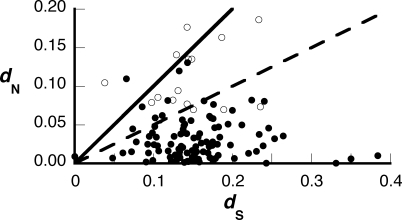
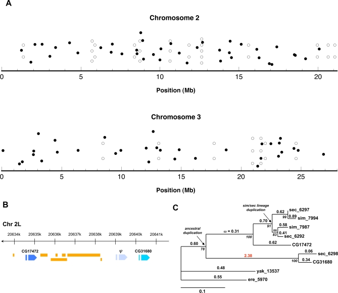
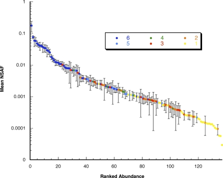
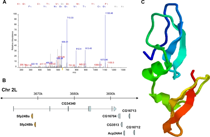
Across diverse taxa, seminal fluid proteins (Sfps) transferred at mating affect the reproductive success of both sexes. Such reproductive proteins often evolve under positive selection between species; because of this rapid divergence, Sfps are hypothesized to play a role in speciation by contributing to reproductive isolation between populations. In Drosophila, individual Sfps have been characterized and are known to alter male sperm competitive ability and female post-mating behavior, but a proteomic-scale view of the transferred Sfps has been missing. Here we describe a novel proteomic method that uses whole-organism isotopic labeling to detect transferred Sfps in mated female D. melanogaster. We identified 63 proteins, which were previously unknown to function in reproduction, and confirmed the transfer of dozens of predicted Sfps. Relative quantification of protein abundance revealed that several of these novel Sfps are abundant in seminal fluid. Positive selection and tandem gene duplication are the prevailing forces of Sfp evolution, and comparative proteomics with additional species revealed lineage-specific changes in seminal fluid content. We also report a proteomic-based gene discovery method that uncovered 19 previously unannotated genes in D. melanogaster. Our results demonstrate an experimental method to identify transferred proteins in any system that is amenable to isotopic labeling, and they underscore the power of combining proteomic and evolutionary analyses to shed light on the complex process of Drosophila reproduction.
Conflict of interest statement
Figures
References
-
- Poiani A. Complexity of seminal fluid: a review. Behav Ecol Sociobiol. 2006;60:289–310.
-
- Clark NL, Aagaard JE, Swanson WJ. Evolution of reproductive proteins from animals and plants. Reproduction. 2006;131:11–22. - PubMed
-
- Swanson WJ, Vacquier VD. Reproductive protein evolution. Annu Rev Ecol Syst. 2002;33:161–179.
-
- Coyne JA, Orr HA. Speciation. Sunderland (Massachusetts): Sinauer Associates; 2004. 545
-
- Martin OY, Hosken DJ. The evolution of reproductive isolation through sexual conflict. Nature. 2003;423:979–982. - PubMed
Publication types
MeSH terms
Substances
Associated data
- Actions
- Actions
- Actions
- Actions
- Actions
- Actions
- Actions
- Actions
- Actions
- Actions
- Actions
- Actions
- Actions
- Actions
- Actions
- Actions
- Actions
- Actions
- Actions
Grants and funding
- HD057974/HD/NICHD NIH HHS/United States
- R01 DK069386/DK/NIDDK NIH HHS/United States
- T32 HG000035/HG/NHGRI NIH HHS/United States
- R01 HD057974/HD/NICHD NIH HHS/United States
- T32 HG00035/HG/NHGRI NIH HHS/United States
- RR011823/RR/NCRR NIH HHS/United States
- HD054631/HD/NICHD NIH HHS/United States
- R01 HD042563/HD/NICHD NIH HHS/United States
- HD42563/HD/NICHD NIH HHS/United States
- P41 RR011823/RR/NCRR NIH HHS/United States
- R56 DK069386/DK/NIDDK NIH HHS/United States
- R03 HD054631/HD/NICHD NIH HHS/United States
- DK069386/DK/NIDDK NIH HHS/United States
LinkOut - more resources
Full Text Sources
Other Literature Sources
Molecular Biology Databases
