. 2008 Jul 30:8:75.
doi: 10.1186/1471-213X-8-75.

Msx1 and Msx2 are required for endothelial-mesenchymal transformation of the atrioventricular cushions and patterning of the atrioventricular myocardium

Affiliations

Msx1 and Msx2 are required for endothelial-mesenchymal transformation of the atrioventricular cushions and patterning of the atrioventricular myocardium

Yi-Hui Chen et al. BMC Dev Biol. .

Abstract

Background: Msx1 and Msx2, which belong to the highly conserved Nk family of homeobox genes, display overlapping expression patterns and redundant functions in multiple tissues and organs during vertebrate development. Msx1 and Msx2 have well-documented roles in mediating epithelial-mesenchymal interactions during organogenesis. Given that both Msx1 and Msx2 are crucial downstream effectors of Bmp signaling, we investigated whether Msx1 and Msx2 are required for the Bmp-induced endothelial-mesenchymal transformation (EMT) during atrioventricular (AV) valve formation.

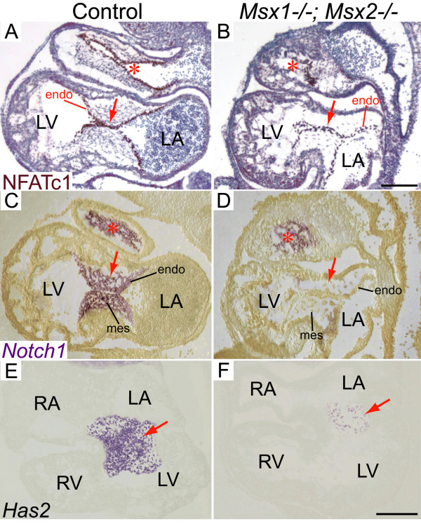
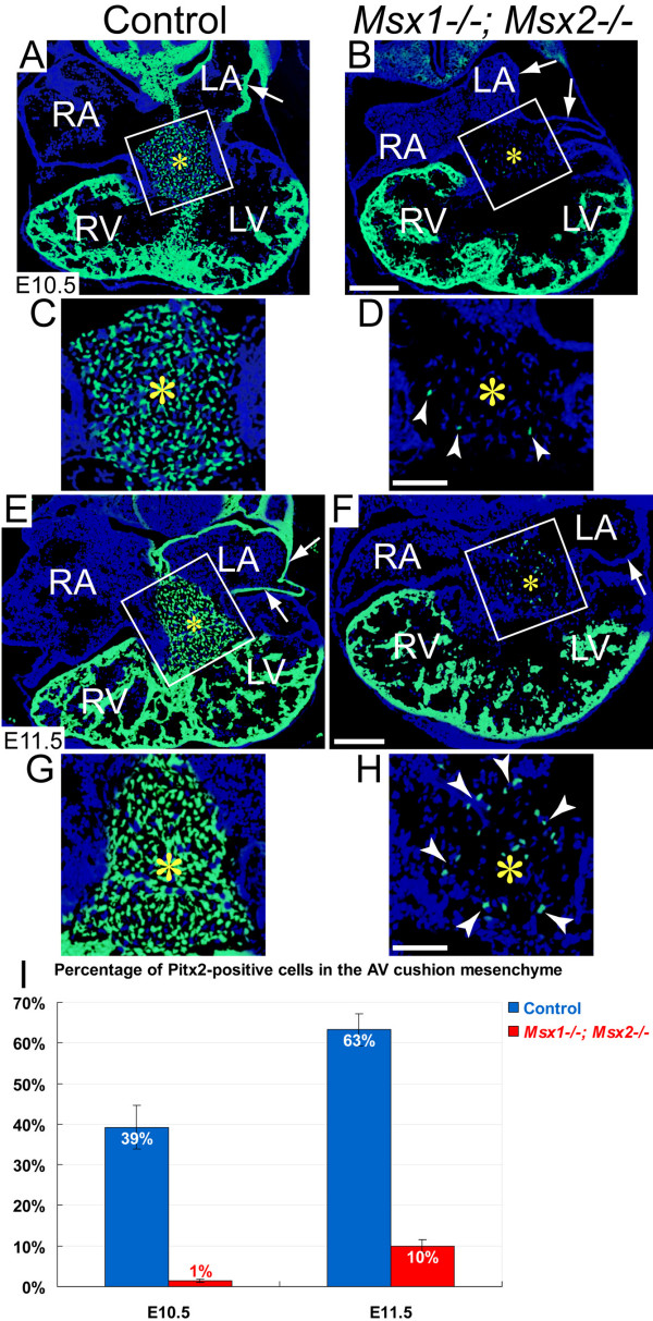
Results: While both Msx1-/- and Msx2-/- single homozygous mutant mice exhibited normal valve formation, we observed hypoplastic AV cushions and malformed AV valves in Msx1-/-; Msx2-/- mutants, indicating redundant functions of Msx1 and Msx2 during AV valve morphogenesis. In Msx1/2 null mutant AV cushions, we found decreased Bmp2/4 and Notch1 signaling as well as reduced expression of Has2, NFATc1 and Notch1, demonstrating impaired endocardial activation and EMT. Moreover, perturbed expression of chamber-specific genes Anf, Tbx2, Hand1 and Hand2 reveals mispatterning of the Msx1/2 double mutant myocardium and suggests functions of Msx1 and Msx2 in regulating myocardial signals required for remodelling AV valves and maintaining an undifferentiated state of the AV myocardium.

Conclusion: Our findings demonstrate redundant roles of Msx1 and Msx2 in regulating signals required for development of the AV myocardium and formation of the AV valves.


Figures

Figure 1
Figure 1
Hypoplastic AV cushions and disorganized AV valves in Msx1-/-; Msx2-/- double mutants at E15.5. Panels D-F are enlarged views of the boxes in panels A-C from histological sections of E15.5 embryonic hearts. Compared with the control (D), Msx1/2 null mutants have compromised AV cushions (indicated by white asterisks in the control and red asterisks in Msx1/2 double mutants), shorter and deformed AV valves extending from the cushions (indicated by triangle arrowheads in panels E and F), and hyperplastic AV valves extending from the AV myocardium (indicated by arrows in panels E and F), suggesting impaired valve outgrowth and remodeling following deficient EMT to form the AV cushions. The red arrow indicates atrial septal defect. IVS, interventricular septum; LA and LV, left atrium and ventricle; RA and RV, right atrium and ventricle. Scale bars: 0.5 mm in A (for A-C) and 0.2 mm in D, and F (for E, F).
Figure 2
Figure 2
Expression of Msx1 and Msx2 in the developing AV canal. Section in situ hybridization showed that the domains of Msx1 and Msx2 expression overlap in a subpopulation of endocardial and cushion mesenchymal cells during AV cushion morphogenesis between E9.5 and E11.5 (A-F). Msx2 also displayed strong expression in the myocardium of the AV canal and right ventricle at all developmental stages examined (B, D and F). Red asterisks indicate AV cushions. endo, endocardium; FG, foregut; IAS and IVS, interatrial and interventricular septum; LA and LV, left atrium and ventricle; myo, myocardium; RA and RV, right atrium and ventricle. Scale bars: 0.1 mm in A (for A, B), 0.2 mm in C (for C, D) and E (for E, F).
Figure 3
Figure 3
Impaired expression of endocardial and cushion mesenchymal genes in Msx1-/-; Msx2-/- mutant AV cushions. At E10.5, the immunostaining intensity of NFATc1 was reduced in Msx1-/-; Msx2-/- mutant AV endocardium compared with the littermate control (red arrows in A, B), while the intensity was comparable between control and Msx1/2 double mutant OFT endocardium (red asterisks in A, B). At the same developmental stage, RNA section in situ hybridization revealed greatly diminished expression of Notch1 (arrows in C, D) in the Msx1/2 double mutant AV cushion mesenchyme and endocardium compared with the control. In contrast, Notch1 expression was normal in the Msx1/2 mutant OFT cushions (compare red asterisks in C and D). Note also a decreased number of Has2-expressing cells in the Msx1/2 null mutant AV cushion mesenchyme (red arrows in E, F). endo, endocardium; LA and LV, left atrium and ventricle; mes, mesenchyme; RA and RV, right atrium and ventricle. Scale bars: 0.1 mm in B (for A-D), 0.2 mm in F (for E, F).
Figure 4
Figure 4
Normal proliferation but decreased total cell numbers in Msx1-/-; Msx2-/- mutant AV cushions during EMT. Immunostaining with the anti-BrdU antibody at both E11.5 and E12.5 revealed comparable levels of cell proliferation in the AV cushions and atrial and ventricular myocardium of Msx1-/-; Msx2-/- mutants and their littermate controls (compare A with B and E with F). Immunostaining against α-smooth muscle actin (α-SMA) showed dramatically reduced numbers of α-SMA-positive mesenchymal cells in the Msx1/2 null mutant AV cushions compared with the control (red arrows in D and H indicate sparse α-SMA-positive cells in the double mutant AV cushions). White asterisks in A, B, E and F, and red asterisks in C, D, G and H indicate the location of the AV cushions. I, Statistical analyses revealed significantly reduced total cell numbers in the AV cushions of the Msx1/2 double mutants compared with those of their littermate controls (at least 4 sections were counted for each embryo, and 3 pairs of Msx1/2 double mutants and their littermate controls were analysed; P < 0.01 for both E10.5 and E11.5, Student's t test). IVS, interventricular septum; LA and LV, left atrium and ventricle; RA and RV, right atrium and ventricle. Scale bars: 0.1 mm in A (for A, B), C (for C, D), F (for E, F) and H (for G, H).
Figure 5
Figure 5
Reduced Bmp2/4 signaling in Msx1-/-; Msx2-/- mutant AV cushions and myocardium during EMT. Panels E-H and M-P are enlarged views of the boxes in panels A-D (at E10.5) and I-L (at E11.5), respectively. Immunostaining for Bmp2/4 (green fluorescence in A, B, E, F, I, J, M and N) and phosphorylated Smad1/5/8 (red fluorescence in C, D, G, H, K, L, O and P) revealed dramatically reduced Bmp2/4-positive and phospho-Smad-positive signals in the Msx1/2 mutant AV cushion mesenchyme (red asterisks in B, F, J, N and yellow asterisks in D, H, L, P) and myocardium (white triangle arrowheads in B, D, J and L) compared with the control (A, C, E, G, I, K, M and O). On the other hand, Msx1-/-; Msx2-/- mutants exhibit broader expression patterns of both Bmp2/4 and Smad1/5/8 in the pharyngeal mesoderm compared with controls (indicated by white arrows in A-D). Q and R indicate average percentages of Bmp2/4-positive and phospho-Smad1/5/8-positive cells in the control and Msx1/2 mutant AV cushions at E10.5 and E11.5 (n = 3 for each developmental stage, P < 0.001, Student's t test). LA and LV, left atrium and ventricle; RA and RV, right atrium and ventricle. Scale bars: 0.1 mm in A (for A-D), F (for E-H), I (for I-L) and N (for M-P).
Figure 6
Figure 6
Perturbed expression of chamber-specific genes in Msx1-/-; Msx2-/- mutant myocardium. RNA section in situ hybridization at E10.5 demonstrated that Tbx2 expression was dramatically reduced in the Msx1/2 null mutant AV myocardium compared with the littermate control (compare the staining intensity in the regions pointed by red notched arrowheads in A and B). In contrast, the level of Tbx expression in the pharyngeal mesoderm was comparable between the control and the Msx1/2 double mutant (indicated by black arrows in A and B). Concomitant with decreased Tbx2 expression in the Msx1-/-; Msx2-/- mutant AV myocardium, Anf expression exhibited increased expression in the Msx1/2 double mutant AV myocardium (compare the staining intensity in the regions pointed by red notched arrowheads in C and D). In addition, we detected ectopic Anf expression in the double mutant right ventricle (red asterisk in D), which was likely a hemodynamic effect secondary to the changes of gene expression in the AV canal. Msx1/2 null mutants also exhibited decreased expression of Hand1 (F) and Hand2 (H) in the AV myocardium (red notched arrowheads). In the Msx1/2 mutant AV myocardium, Hand1 expression was almost undetectable and Hand2 expression was significantly reduced compared with the control (red notched arrowheads in E-H). LA and LV, left atrium and ventricle; RA and RV, right atrium and ventricle. Scale bars: 0.1 mm in A (for A-D) and E (for E-H).
Figure 7
Figure 7
Decreased contribution of Pitx2-positive cells to the Msx1-/-; Msx2-/- mutant AV cushion mesenchyme. Panels C, D, G and H are enlarged views of the boxes in A, B (at E10.5) and E, F (at E11.5), respectively. Pitx2 immunostaining (green fluorescence) revealed a substantial reduction in the number of Pitx2-positive cells in the Msx1-/-; Msx2-/- mutant AV cushion mesenchyme compared with the control (compare the regions marked by yellow asterisks in C, D, G and H). White notched arrowheads in D and H indicate sparse Pitx2-positive mesenchymal cells in the Msx1/2 null mutant AV cushions. There was no detectable Pitx2 expression in the left atrial myocardium of the Msx1/2 double mutants shown in B and F (indicated by white arrows). I, Statistical analyses revealed significantly reduced percentages of Pitx2-positive cells in the AV cushion mesenchyme of the Msx1/2 double mutants at both E10.5 and E11.5 (n = 3 for each developmental stage, P < 0.01 at E10.5, P < 0.001 at E11.5, Student's t test). LA and LV, left atrium and ventricle; RA and RV, right atrium and ventricle. Scale bars: 0.1 mm in B (for A, B) and F (for E, F), 0.05 mm in D (for C, D) and H (for G, H).

Similar articles

Cited by

References

    1. Schroeder JA, Jackson LF, Lee DC, Camenisch TD. Form and function of developing heart valves: coordination by extracellular matrix and growth factor signaling. J Mol Med. 2003;81:392–403. - PubMed
    1. Srivastava D, Olson EN. A genetic blueprint for cardiac development. Nature. 2000;407:221–226. - PubMed
    1. Zaffran S, Frasch M. Early signals in cardiac development. Circ Res. 2002;91:457–469. - PubMed
    1. Armstrong EJ, Bischoff J. Heart valve development. Endothelial cell signaling and differentiation. Circ Res. 2004;95:459–470. - PMC - PubMed
    1. Eisenberg LM, Markwald RR. Molecular regulation of atrioventricular valvuloseptal morphogenesis. Circ Res. 1995;77:1–6. - PubMed

MeSH terms