

Transcriptional profiling of putative human epithelial stem cells
- PMID: 18667080
- PMCID: PMC2536675
- DOI: 10.1186/1471-2164-9-359
Transcriptional profiling of putative human epithelial stem cells
Abstract
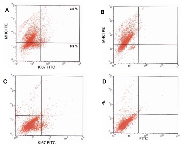
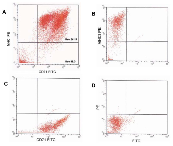
Background: Human interfollicular epidermis is sustained by the proliferation of stem cells and their progeny, transient amplifying cells. Molecular characterization of these two cell populations is essential for better understanding of self renewal, differentiation and mechanisms of skin pathogenesis. The purpose of this study was to obtain gene expression profiles of alpha 6+/MHCI+, transient amplifying cells and alpha 6+/MHCI-, putative stem cells, and to compare them with existing data bases of gene expression profiles of hair follicle stem cells. The expression of Major Histocompatibility Complex (MHC) class I, previously shown to be absent in stem cells in several tissues, and alpha 6 integrin were used to isolate MHCI positive basal cells, and MHCI low/negative basal cells.
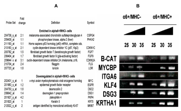
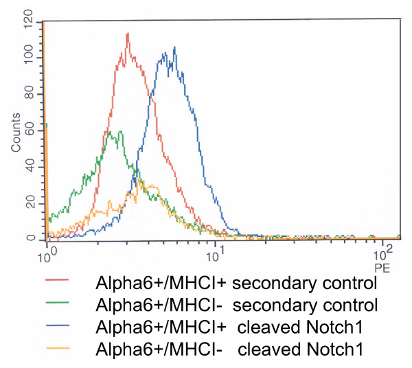
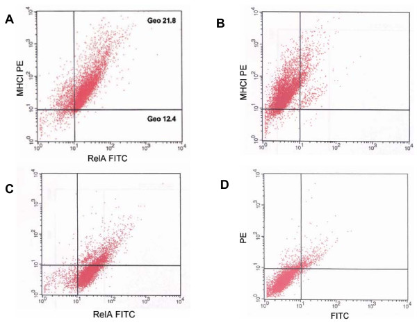
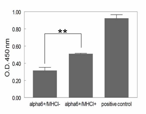
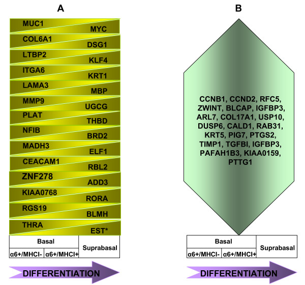
Results: Transcriptional profiles of the two cell populations were determined and comparisons made with published data for hair follicle stem cell gene expression profiles. We demonstrate that presumptive interfollicular stem cells, alpha 6+/MHCI- cells, are enriched in messenger RNAs encoding surface receptors, cell adhesion molecules, extracellular matrix proteins, transcripts encoding members of IFN-alpha family proteins and components of IFN signaling, but contain lower levels of transcripts encoding proteins which take part in energy metabolism, cell cycle, ribosome biosynthesis, splicing, protein translation, degradation, DNA replication, repair, and chromosome remodeling. Furthermore, our data indicate that the cell signaling pathways Notch1 and NF-kappaB are downregulated/inhibited in MHC negative basal cells.
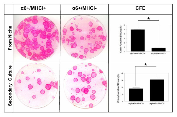
Conclusion: This study demonstrates that alpha 6+/MHCI- cells have additional characteristics attributed to stem cells. Moreover, the transcription profile of alpha 6+/MHCI- cells shows similarities to transcription profiles of mouse hair follicle bulge cells known to be enriched for stem cells. Collectively, our data suggests that alpha 6+/MHCI- cells may be enriched for stem cells. This study is the first comprehensive gene expression profile of putative human epithelial stem cells and their progeny that were isolated directly from neonatal foreskin tissue. Our study is important for understanding self renewal and differentiation of epidermal stem cells, and for elucidating signaling pathways involved in those processes. The generated data base may serve those working with other human epithelial tissue progenitors.
Figures
Similar articles
-
Location and phenotype of human adult keratinocyte stem cells of the skin.Differentiation. 2004 Oct;72(8):387-95. doi: 10.1111/j.1432-0436.2004.07208005.x. Differentiation. 2004. PMID: 15606498
-
Brg1 governs a positive feedback circuit in the hair follicle for tissue regeneration and repair.Dev Cell. 2013 Apr 29;25(2):169-81. doi: 10.1016/j.devcel.2013.03.015. Epub 2013 Apr 18. Dev Cell. 2013. PMID: 23602386
-
Capturing and profiling adult hair follicle stem cells.Nat Biotechnol. 2004 Apr;22(4):411-7. doi: 10.1038/nbt950. Epub 2004 Mar 14. Nat Biotechnol. 2004. PMID: 15024388
-
Epithelial stem cells and implications for wound repair.Semin Cell Dev Biol. 2012 Dec;23(9):946-53. doi: 10.1016/j.semcdb.2012.10.001. Epub 2012 Oct 17. Semin Cell Dev Biol. 2012. PMID: 23085626 Free PMC article. Review.
-
Defining Transcriptional Signatures of Human Hair Follicle Cell States.J Invest Dermatol. 2020 Apr;140(4):764-773.e4. doi: 10.1016/j.jid.2019.07.726. Epub 2019 Oct 31. J Invest Dermatol. 2020. PMID: 31676413 Free PMC article. Review.
Cited by
-
Comparative transcriptional profiling of the limbal epithelial crypt demonstrates its putative stem cell niche characteristics.BMC Genomics. 2010 Sep 29;11:526. doi: 10.1186/1471-2164-11-526. BMC Genomics. 2010. PMID: 20920242 Free PMC article.
-
Restoration of Immune Privilege in Human Dermal Papillae Controlling Epithelial-Mesenchymal Interactions in Hair Formation.Tissue Eng Regen Med. 2022 Feb;19(1):105-116. doi: 10.1007/s13770-021-00392-7. Epub 2021 Oct 9. Tissue Eng Regen Med. 2022. PMID: 34626334 Free PMC article.
-
Frequent somatic reversion of KRT1 mutations in ichthyosis with confetti.J Clin Invest. 2015 Apr;125(4):1703-7. doi: 10.1172/JCI64415. Epub 2015 Mar 16. J Clin Invest. 2015. PMID: 25774499 Free PMC article.
-
Immunogenicity and tolerance induction in vascularized composite allotransplantation.Front Transplant. 2024 Feb 13;3:1350546. doi: 10.3389/frtra.2024.1350546. eCollection 2024. Front Transplant. 2024. PMID: 38993748 Free PMC article. Review.
-
Annotation enrichment analysis: an alternative method for evaluating the functional properties of gene sets.Sci Rep. 2014 Feb 26;4:4191. doi: 10.1038/srep04191. Sci Rep. 2014. PMID: 24569707 Free PMC article.
References
-
- Bickenbach JR, McCutecheon J, Mackenzie IC. Rate of loss of tritiated thymidine label in basal cells in mouse epithelial tissues. Cell Tissue Kinet. 1986;19:325–333. - PubMed
-
- Bickenbach JR. Identification and behavior of label-retaining cells in oral mucosa and skin. J Dent Res. 1981;60:1611–1620. - PubMed
Publication types
MeSH terms
Substances
Grants and funding
LinkOut - more resources
Full Text Sources
Other Literature Sources
Medical
Molecular Biology Databases
Research Materials
