Comparative Study
. 2008 Jul 31;59(2):226-40.
doi: 10.1016/j.neuron.2008.06.025.

Coordinated actions of the forkhead protein Foxp1 and Hox proteins in the columnar organization of spinal motor neurons

Affiliations
Comparative Study

Coordinated actions of the forkhead protein Foxp1 and Hox proteins in the columnar organization of spinal motor neurons

David L Rousso et al. Neuron. .

Abstract

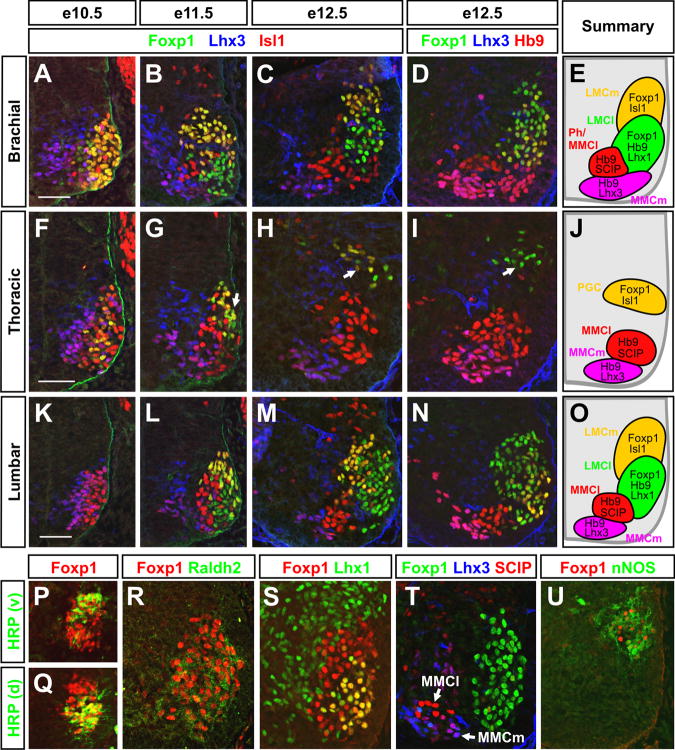
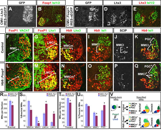
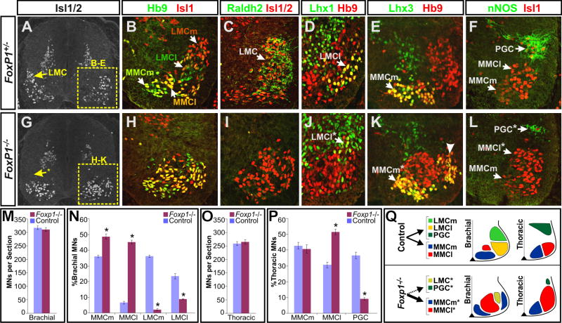
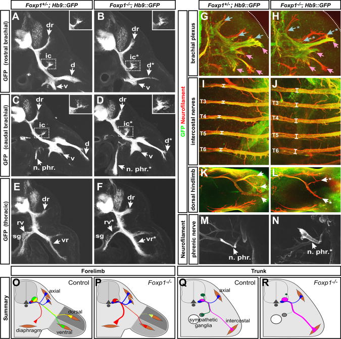
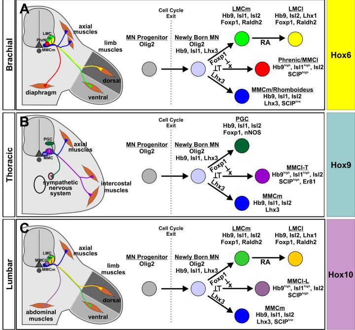
The formation of locomotor circuits depends on the spatially organized generation of motor columns that innervate distinct muscle and autonomic nervous system targets along the body axis. Within each spinal segment, multiple motor neuron classes arise from a common progenitor population; however, the mechanisms underlying their diversification remain poorly understood. Here, we show that the Forkhead domain transcription factor Foxp1 plays a critical role in defining the columnar identity of motor neurons at each axial position. Using genetic manipulations, we demonstrate that Foxp1 establishes the pattern of LIM-HD protein expression and accordingly organizes motor axon projections, their connectivity with peripheral targets, and the establishment of motor pools. These functions of Foxp1 act in accordance with the rostrocaudal pattern provided by Hox proteins along the length of the spinal cord, suggesting a model by which motor neuron diversity is achieved through the coordinated actions of Foxp1 and Hox proteins.


Figures

Figure 1
Figure 1. Foxp1 is selectively expressed by developing LMC and PGC MNs and distinguishes these cells from MMCm and MMCl MNs
(A-O) Antibody costaining analysis of Foxp1 and LIM-HD protein expression in the developing mouse spinal cord. (P-Q) HRP injections into ventral (P) or dorsal (Q) limb muscles at e13.5 confirms that Foxp1 is present in both LMC MN populations. (R-T) Foxp1 expression in the e12.5 rostral brachial spinal cord coincides with the expression of the LMC markers Raldh2 and Lhx1 but not SCIP, which is expressed by MMCm and MMCl MNs. (U) Foxp1 expression in the e12.5 thoracic spinal cord coincides with the PGC MN marker nNOS. Ventrolateral quadrants of the spinal cord are shown in all images. Scale bars = 50 μm.
Figure 2
Figure 2. Foxp1 misexpression is sufficient to repress Lhx3 and MMC MN fates, and promotes the early formation of LMC and PGC MNs
(A-E) Effects of misexpression of Lhx3 or Foxp1 in the brachial spinal cord of chick embryos. Images representative of 5-10 embryos for each experiment. (F, L) Costaining analysis of Foxp1 and the general MN marker VAChT demonstrates that Foxp1 is expressed by most MNs in e11.5 Hb9::Foxp1 transgenic animals. Arrows indicate the normal position of MMC MNs in control and transgenic animals. (G-J; M-P) Analysis of the rostral forelimbs reveals an expansion in the production of Lhx1+ LMCl MNs, and a reduction in both Lhx3+ MMCm (H, N), and Isl1high Hb9+ SCIPhigh MMCl MNs (I, J, O, P). (K,Q) The transgenic misexpression of Foxp1 at thoracic levels increases the appearance of dorsally migrating Isl1+ Hb9low PGC MNs, and decreases the number of Isl1high Hb9+ MMCl MNs. (R-U) Quantification of MN numbers in Hb9::Foxp1 and littermate control embryos. Mean ± SEM were calculated by pooling multiple sections collected from at least two embryos of each genotype. Results are representative of 12 embryos analyzed. (R, T) Hb9::Foxp1 animals show a small reduction in total MNs at brachial and thoracic levels, p < 0.001 and p < 0.01 respectively. (S) MMCm and MMCl MNs were reduced in Hb9::Foxp1 embryos (p < 0.001 in both cases), while LMCm and LMCl MNs were increased (p < 0.05 and p < 0.001, respectively). (U) Foxp1 misexpression at thoracic levels led to a small decrease in MMCm formation (p = 0.16), a more significant decrease in MMCl formation (p < 0.05), and an increase in PGC MN formation (p < 0.001). (V) Schematic summary of the misexpression experiments.
Figure 3
Figure 3. LMC and PGC MNs are transformed into MMC MNs in the absence of Foxp1
(A-L) Antibody costaining analysis of transverse sections of e12.5 FoxP1+/- heterozygous and FoxP1-/- homozygous mutant littermates indicates an alteration in spinal motor column identities. (B, C, D, H, I, J) Analysis of LIM-HD protein and Raldh2 expression at brachial levels indicates a considerable loss of LMC motor neurons in Foxp1-null animals, and an increased genesis of Hb9+ Isl1high MMCl MNs. (E, K) Foxp1 mutants show an excessive production of laterally positioned MMCm-like cells that resemble rhomboideus MNs. (F,L) nNOS staining at thoracic levels indicates a dramatic loss of sympathetic MNs in the Foxp1 mutant mice, and a corresponding increase in the generation of Isl1+ MMCl MNs. (M-O) Quantification of MN numbers in e12.5 Foxp1-/- mutants and littermate controls. Mean ± SEM were calculated by pooling multiple sections collected from at least two embryos of each genotype. Motor column identities were designated by the following antibody costaining combinations: MMCm, Lhx3+ Isl1+; MMCl, Lhx3- Hb9+ Isl1high; LMCm, Hb9low Isl1+; LMCl, Lhx1+ Hb9+; PGC, nNOS+ Isl1+. (M) Total MN numbers are not significantly changed in the Foxp1 mutants. (N) Analysis in the mid-forelimb level (C5-C7) shows a significant increase in the generation of MMCm and MMCl MNs and loss of LMCm and LMCl MNs (p < 0.0001 in all cases). (O) Analysis of motor column distribution at thoracic levels shows no change in MMCm MN formation, but an increased generation of MMCl MNs and concomitant loss of PGC MNs in the Foxp1 mutants (p < 0.0001 in both cases). (Q) Schematic summary of the Foxp1 mutant phenotype.
Figure 4
Figure 4. Redirection of LMC and PGC motor fibers toward MMC muscle targets in Foxp1 mutants
(A-F) Analysis of motor fibers at e11.5 in vibratome sections of Hb9::GFP; Foxp1-null and littermate control embryos. Sections represent the following positions: (A, B) rostral brachial plexus, (C, D) caudal brachial plexus, and (E, F) rostral thoracic level. dr, dorsal ramus; ic, ramus intercostalis externus; d, dorsal plexus; n. phr., phrenic nerve; v, ventral plexus; rv, ramus visceralis; vr, ventral ramus; sg, sympathetic ganglia. (G-N). Whole mount immunohistochemistry of motor (Hb9::GFP, green) and sensory plus motor fibers (neurofilament, red) in (G, H) the e11.5 brachial plexus, (I, J) e12.5 rostral intercostal nerves, (K, L) e12.5 dorsal hindlimb, and (M, N) e12.5 phrenic nerve (M, N). Blue and pink arrows in (G-H) designates dorsally and ventrally projecting nerves, respectively. Intercostal nerves in (I, J) were found to have an average diameter of 28.3±1.0 μm in control embryos and 40.3±1.9 μm in Foxp1 mutants, p < 0.01. * in all panels designates locations with reproducible changes in axon projections. Images are representative of > 3 embryos of each genotype analyzed. (O-R) Schematic summary of axon misprojections.
Figure 5
Figure 5. Topographic misprojections of motor axons in Foxp1 mutant embryos
(A-D). MN projections to axial muscles were traced using HRP injections into axial muscles in e13.5 control and Foxp1 mutant embryos, and subjected to costaining analysis with the indicted antibodies. (E-H) MN projections to the dorsal limbs were similarly traced using HRP injections. In both control and Foxp1 mutants, most dorsal projecting MNs lacked Isl1 staining and instead expressed Lhx1 (data not shown). However, in the Foxp1 mutants, some of the dorsally projecting neurons aberrantly expressed Isl1 (inset in panel F). (I-L) Injections of HRP into the ventral limbs labels a dorsolaterally positioned group of Isl1+ cells in the controls, and a ventromedially positioned group of Isl1+ MN in Foxp1 mutants. Some ventrally projecting MNs in the Foxp1 mutants express both Lhx1 and Isl1 (inset in panel J). Arrows in panels J and L indicate the unusual horizontal morphology of dendrites labeled by retrograde labeling from the ventral limbs in the Foxp1 mutants. (M) Quantification of retrograde labeling of MNs following HRP injections into dorsal and ventral limb muscles. The percentage of HRP labeled MNs that are Isl1+ following injections into the dorsal limb or Lhx1+ following injections into the ventral limbs are shown (p < 0.05 in both cases). (N, O, Q, R) Distribution of EphA4 receptor in vibratome sections of the rostral brachial plexus from control or Foxp1-null littermates. (P, S) Equivalent analysis of EphA4 expression in thoracic sections of wild-type embryos. SG, sympathetic ganglia; IC, intercostal nerves.
Figure 6
Figure 6. Foxp1 is required for the appropriate formation of LMC-associated motor pools
(A-H) Antibody costaining analysis of motor pool markers in the e13.0-e13.5 rostral hindlimb. Images representative of > 4 embryos of each genotype analyzed.
Figure 7
Figure 7. Cooperative functions of Foxp1 and Hox proteins are required for the formation of LMC MNs and motor pools
(A-H) Antibody costaining analysis of lumbar (L1-L2) sections of e13.5 Hoxa10+/-; Hoxc10+/-; Hoxd10+/- 3-allele control and Hoxa10+/-; Hoxc10-/-; Hoxd10-/- 5-allele mutant embryos reveals changes in the pattern of Foxp1 expression and its abnormal association with the PGC marker nNOS rather than the LMCl marker Lhx1 and the LMCm motor pool marker Er81. Hox10 5-allele mutants also show an expansion of thoracic MMCl MNs, which express Er81 (MMCl-T), into the lumbar spinal cord. (I-J) Analysis of motor column formation in the lumbar spinal cord (L1-L2) of age-matched Foxp1 mutant embryos. Foxp1 mutants show a reduced formation of LMCl MNs and an increased formation of MMCl that lack Er81 expression (MMCl-L). (M-N) Distribution of Foxp1+ MNs as LMC-associated (nNOS-) and PGC-associated (nNOS+) along the rostrocaudal extent of the lumbar spinal cord of Hox10 3-allele control and Hox10 5-allele mutant litermates. Counts are representative of 3 embryos examined per genotype. (O) Summary of the coordinate functions of Hox10 and Foxp1 in the determination of thoracic and lumbar motor columns and motor pools. In Hox10 5-allele mutants LMC MNs are transformed to a PGC fate, and lumbar MMCl MNs express the thoracic motor pool marker Er81 (MMCl-T). In Foxp1 mutants, LMC MNs are transformed into lumbar MMCl MNs that lack Er81 expression (MMCl-L). LMC MNs and LMC-associated motor pools only form in the presence of both Hoxc10/d10 and Foxp1.
Figure 8
Figure 8. The integrated functions of Foxp1 and Hox proteins determine the columnar fate of MNs throughout the body
(A-C) Proposed models for how different classes of MNs are formed at distinct rostrocaudal positions. At each axial level, MNs arise from a common population of Olig2+ neural progenitors. Soon after cell cycle exit, newly born MNs adopt one of three potential fates due to cross-repressive interactions between Lhx3 and Foxp1, and the ability of Foxp1 to block MMCl MN development. MMCl MN formation may further depend upon the function of an additional determinant (X). The establishment of segment-specific motor columns and motor pools then proceeds in accordance to the Hox protein profile expressed by the MNs. Hox proteins may further participate in the Foxp1-dependent intrasegmental patterning of MNs by regulating the level of its expression. LMC MN diversification is further driven by the actions of retinoid signaling (Sockanathan and Jessell, 1998; Sockanathan et al., 2003).

References

    1. Arber S, Han B, Mendelsohn M, Smith M, Jessell TM, Sockanathan S. Requirement for the homeobox gene Hb9 in the consolidation of motor neuron identity. Neuron. 1999;23:659–674. - PubMed
    1. Arber S, Ladle DR, Lin JH, Frank E, Jessell TM. ETS gene Er81 controls the formation of functional connections between group Ia sensory afferents and motor neurons. Cell. 2000;101:485–498. - PubMed
    1. Briscoe J, Novitch BG. Regulatory pathways linking progenitor patterning, cell fates and neurogenesis in the ventral neural tube. Philos Trans R Soc Lond B Biol Sci. 2008;363:57–70. - PMC - PubMed
    1. Butler SJ, Tear G. Getting axons onto the right path: the role of transcription factors in axon guidance. Development. 2007;134:439–448. - PubMed
    1. Carpenter EM, Goddard JM, Davis AP, Nguyen TP, Capecchi MR. Targeted disruption of Hoxd-10 affects mouse hindlimb development. Development. 1997;124:4505–4514. - PubMed

Publication types