. 2008 Oct;82(19):9739-52.
doi: 10.1128/JVI.00871-08. Epub 2008 Jul 30.

Potent antibody-mediated neutralization and evolution of antigenic escape variants of simian immunodeficiency virus strain SIVmac239 in vivo

Affiliations

Potent antibody-mediated neutralization and evolution of antigenic escape variants of simian immunodeficiency virus strain SIVmac239 in vivo

Shuji Sato et al. J Virol. 2008 Oct.

Abstract

Here, we describe the evolution of antigenic escape variants in a rhesus macaque that developed unusually high neutralizing antibody titers to SIVmac239. By 42 weeks postinfection, 50% neutralization of SIVmac239 was achieved with plasma dilutions of 1:1,000. Testing of purified immunoglobulin confirmed that the neutralizing activity was antibody mediated. Despite the potency of the neutralizing antibody response, the animal displayed a typical viral load profile and progressed to terminal AIDS with a normal time course. Viral envelope sequences from week 16 and week 42 plasma contained an excess of nonsynonymous substitutions, predominantly in V1 and V4, including individual sites with ratios of nonsynonymous to synonymous substitution rates (dN/dS) highly suggestive of strong positive selection. Recombinant viruses encoding envelope sequences isolated from these time points remained resistant to neutralization by all longitudinal plasma samples, revealing the failure of the animal to mount secondary responses to the escaped variants. Substitutions at two sites with significant dN/dS values, one in V1 and one in V4, were independently sufficient to confer nearly complete resistance to neutralization. Substitutions at three additional sites, one in V4 and two in gp41, conferred moderate to high levels of resistance when tested individually. All the amino acid changes leading to escape resulted from single nucleotide substitutions. The observation that antigenic escape resulted from individual, single amino acid replacements at sites well separated in current structural models of Env indicates that the virus can utilize multiple independent pathways to rapidly achieve similar levels of resistance.


Figures

FIG. 1.
FIG. 1.
Viral replication in animal Mm333. Viral RNA loads were measured from longitudinal plasma samples (at 0, 1, 2, 3, 4, 6, 12, 16, 20, 26, and 34 weeks postinoculation) taken from animal Mm333 using quantitative real-time RT-PCR. Mean acute peak height and mean post-acute SIV RNA load values (indicated by dotted lines) for SIVmac239-infected rhesus macaques were obtained from a previous study (16) and are included for the purpose of comparison.
FIG. 2.
FIG. 2.
Development of neutralizing plasma titers against SIVmac239 in Mm333. Plasma samples were tested for their ability to neutralize SIVmac239 using C8166-SEAP indicator cells. (A) Neutralization profile of week 42 plasma from four SIVmac239-positive rhesus macaques and a pool of plasma (week 20) from multiple SIVmac239-positive animals. (B) Neutralization profile of Mm333 for longitudinal plasma samples harvested at day of inoculation and at weeks 4, 16, 20, 42, and 58 postinfection, as indicated. All samples were tested in duplicate.
FIG. 3.
FIG. 3.
Neutralization of SIVmac239 by Mm333 plasma is antibody mediated. The neutralization profiles of plasma and purified (purif) total IgG fractions are compared for Mm333 (week 82 postinoculation) and an uninfected macaque. Total IgG was purified from plasma using protein A/G beads. The purified IgG fraction volume was reconstituted with 1× PBS to be equivalent to the starting plasma sample volume so that the neutralizing titers of plasma and purified IgG could be directly compared. All samples were tested in duplicate. wk, week.
FIG. 4.
FIG. 4.
Profile of Mm333 plasma binding to overlapping 15-mer SIVmac239 Env peptides by ELISA. (A) Binding profile of the week 20 plasma pool from multiple SIVmac239-positive, nonneutralizing animals. (B) Binding profile of week 26 Mm333 plasma. The numbers above the bars indicate peptides to which binding of fivefold above background was observed. OD, optical density; TM, transmembrane.
FIG. 5.
FIG. 5.
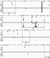
Amino acid sequence alignment of envelope clones isolated from Mm333 week 16 and week 42 plasma. The full-length env coding region was amplified and cloned from plasma viral RNA by RT-PCR, followed by TOPO-TA cloning. Ten individual envelope clones from week 16 plasma and seven from week 42 plasma were sequenced, and variations from the parental SIVmac239 amino acid sequence are shown as white letters within black boxes. The vertical dotted line indicates the cleavage site that separates gp120 and gp41. The leader peptide (LP), variable domains V1 through V5, heptad repeats 1 and 2 (HR1 and HR2), and the transmembrane domain (TM) are outlined and indicated under each region. N-linked glycosylation motifs [NX(S/T)] in the parental sequence are boxed. Positively selected residues identified by dN/dS analysis are indicated by asterisks.
FIG. 5.
FIG. 5.
Amino acid sequence alignment of envelope clones isolated from Mm333 week 16 and week 42 plasma. The full-length env coding region was amplified and cloned from plasma viral RNA by RT-PCR, followed by TOPO-TA cloning. Ten individual envelope clones from week 16 plasma and seven from week 42 plasma were sequenced, and variations from the parental SIVmac239 amino acid sequence are shown as white letters within black boxes. The vertical dotted line indicates the cleavage site that separates gp120 and gp41. The leader peptide (LP), variable domains V1 through V5, heptad repeats 1 and 2 (HR1 and HR2), and the transmembrane domain (TM) are outlined and indicated under each region. N-linked glycosylation motifs [NX(S/T)] in the parental sequence are boxed. Positively selected residues identified by dN/dS analysis are indicated by asterisks.
FIG. 6.
FIG. 6.
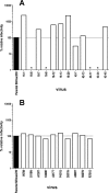
Relative infectivity of SIVmac239 env variants. Infectivity of viruses with week 16 and week 42 full-length envelope clones (sequences are shown in Fig. 5) (A) and single amino acid substitutions (B) were assessed in C8166-SEAP cells. Clones containing stop codons in the env open reading frame were excluded. The percent relative infectivity was calculated as the percentage of the reciprocal of the amount of p27 (in nanograms) of variant virus that gives the same SEAP activity as 1 ng of parental SIVmac239 (100%), determined by infecting the reporter cell line with serially diluted stocks of each virus. An asterisk indicates that SEAP activity above background (uninfected cells) was not detected at the highest concentration of the particular virus tested.
FIG. 7.
FIG. 7.
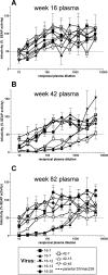
Neutralization resistance of week 16 and week 42 env clones to longitudinal Mm333 plasma samples. SIVmac239 variants encoding env sequences isolated from week 16 (clones 16-1, 16-7, 16-12, 16-13, and 16-20) and week 42 (clones 42-7, 42-13, and 42-45) were tested for neutralization resistance to Mm333 plasma from week 16, week 42, and week 82 using C8166-SEAP cells. All samples were tested in duplicate.
FIG. 8.
FIG. 8.
Neutralization escape imparted by single substitutions in Env. Single residue substitutions in Env identified in neutralization escape clones isolated from Mm333 week 16 and week 42 plasma were introduced in parental SIVmac239 and tested for neutralization resistance to week 82 plasma. (A) Substitutions that increased neutralization resistance to Mm333 plasma. (B) Substitutions that did not affect neutralization resistance. All samples were tested in duplicate.

References

    1. Alexander, L., L. Denekamp, S. Czajak, and R. C. Desrosiers. 2001. Suboptimal nucleotides in the infectious, pathogenic simian immunodeficiency virus clone SIVmac239. J. Virol. 754019-4022. - PMC - PubMed
    1. Anisimova, M., J. P. Bielawski, and Z. Yang. 2002. Accuracy and power of Bayes prediction of amino acid sites under positive selection. Mol. Biol. Evol. 19950-958. - PubMed
    1. Burns, D. P., C. Collignon, and R. C. Desrosiers. 1993. Simian immunodeficiency virus mutants resistant to serum neutralization arise during persistent infection of rhesus monkeys. J. Virol. 674101-4113. - PMC - PubMed
    1. Burns, D. P., and R. C. Desrosiers. 1991. Selection of genetic variants of simian immunodeficiency virus in persistently infected rhesus monkeys. J. Virol. 651843-1854. - PMC - PubMed
    1. Chackerian, B., L. M. Rudensey, and J. Overbaugh. 1997. Specific N-linked and O-linked glycosylation modifications in the envelope V1 domain of simian immunodeficiency virus variants that evolve in the host alter recognition by neutralizing antibodies. J. Virol. 717719-7727. - PMC - PubMed

Publication types