

doi: 10.1128/JVI.00895-08.
Epub 2008 Jul 30.
Structure of flexible filamentous plant viruses
Affiliations
- PMID: 18667514
- PMCID: PMC2546986
- DOI: 10.1128/JVI.00895-08
Item in Clipboard
Structure of flexible filamentous plant viruses
J Virol.
2008 Oct.
Abstract
Flexible filamentous viruses make up a large fraction of the known plant viruses, but in comparison with those of other viruses, very little is known about their structures. We have used fiber diffraction, cryo-electron microscopy, and scanning transmission electron microscopy to determine the symmetry of a potyvirus, soybean mosaic virus; to confirm the symmetry of a potexvirus, potato virus X; and to determine the low-resolution structures of both viruses. We conclude that these viruses and, by implication, most or all flexible filamentous plant viruses share a common coat protein fold and helical symmetry, with slightly less than 9 subunits per helical turn.
Figures
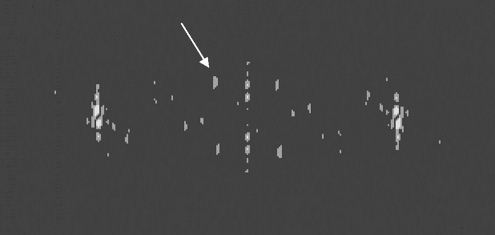
Example of a calculated diffraction pattern from a segment of SMV in a cryo-electron micrograph. The arrow indicates the first intensity peak in the first layer line. The position of this layer line corresponds to a spacing of about 165 Å; near-meridional layer lines corresponding to a spacing of 33 Å are also clearly visible. For phases to be used in symmetry determination, the first-layer-line intensities in the four quadrants were required to be clearly visible and symmetric in position and appearance.
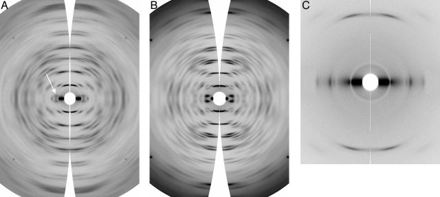
(A) and (B) Wide-angle fiber diffraction patterns from the potyvirus SMV (A) and the potexvirus NMV (B), with intensities corrected and data transformed into reciprocal space. The arrow in panel A indicates the first layer line. (C) Low-angle data from SMV, corrected and transformed.

SDS-PAGE from SMV. Two bands from degradation products of SMV coat protein are clearly visible.
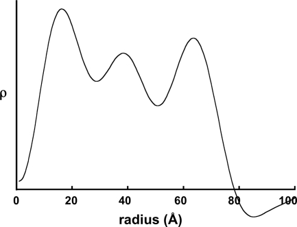
Radial density distribution in SMV. Units of electron density (ρ) are arbitrary.
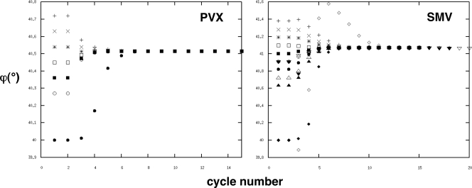
Convergence of the rotation angle ϕ as a function of cycle number during IHRSR. Refinements were started from many different rotation angles; provided that the angle was not too far from the final refined angle (21), they all converged to the same value. Most of the refinements used solid cylinders as initial reference volumes, but for SMV, arbitrary unrelated density distributions (solid inverted triangles, open diamonds, and solid diamonds) were also used.
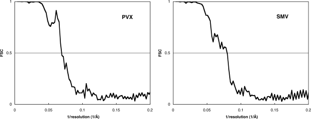
Fourier shell correlation plots for the refined PVX and SMV models. In both cases, the correlations fall below 0.5 at a resolution of about 1/14 Å.
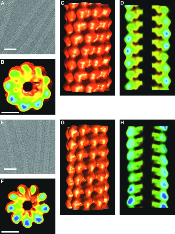
(A) Cryo-electron micrograph of SMV with contrast reversed. Scale bar, 250 Å. (B) IHRSR reconstruction of SMV, section normal to viral axis. Scale bar (also applies to panels C and D), 50 Å. (C) IHRSR reconstruction of SMV, outside surface view. (D) IHRSR reconstruction of SMV, section through viral axis. (E) Cryo-electron micrograph of PVX with contrast reversed. Scale bar, 250 Å. (F) IHRSR reconstruction of PVX, section normal to viral axis. Scale bar (also applies to panels G and H), 50 Å. (G) IHRSR reconstruction of PVX, outside surface view. (H) IHRSR reconstruction of PVX, section through viral axis. Color coding in panels B, C, D, F, G, and H is from red-orange (low density) to green-blue (high density).
Similar articles
-
A common structure for the potexviruses.Virology. 2013 Feb 5;436(1):173-8. doi: 10.1016/j.virol.2012.11.008. Epub 2012 Dec 11. Virology. 2013. PMID: 23245732
-
Atomic structure of potato virus X, the prototype of the Alphaflexiviridae family.Nat Chem Biol. 2020 May;16(5):564-569. doi: 10.1038/s41589-020-0502-4. Epub 2020 Mar 16. Nat Chem Biol. 2020. PMID: 32203412
-
Potyvirus virion structure shows conserved protein fold and RNA binding site in ssRNA viruses.Sci Adv. 2017 Sep 20;3(9):eaao2182. doi: 10.1126/sciadv.aao2182. eCollection 2017 Sep. Sci Adv. 2017. PMID: 28948231 Free PMC article.
-
[Investigation of helical plant virus ribonucleoprotein structures with the help of tritium planigraphy and theoretical modeling].Mol Biol (Mosk). 2004 Sep-Oct;38(5):945-58. Mol Biol (Mosk). 2004. PMID: 15554196 Review. Russian.
-
Helical viruses.Adv Exp Med Biol. 2012;726:631-58. doi: 10.1007/978-1-4614-0980-9_28. Adv Exp Med Biol. 2012. PMID: 22297534 Review.
Cited by
-
The Phylogeography of Potato Virus X Shows the Fingerprints of Its Human Vector.Viruses. 2021 Apr 9;13(4):644. doi: 10.3390/v13040644. Viruses. 2021. PMID: 33918611 Free PMC article.
-
Plug-and-Display Photo-Switchable Systems on Plant Virus Nanoparticles.BioTech (Basel). 2022 Oct 21;11(4):49. doi: 10.3390/biotech11040049. BioTech (Basel). 2022. PMID: 36278561 Free PMC article.
-
Applications of viral nanoparticles in medicine.Curr Opin Biotechnol. 2011 Dec;22(6):901-8. doi: 10.1016/j.copbio.2011.04.020. Epub 2011 May 16. Curr Opin Biotechnol. 2011. PMID: 21592772 Free PMC article. Review.
-
Structural basis for the multitasking nature of the potato virus Y coat protein.Sci Adv. 2019 Jul 17;5(7):eaaw3808. doi: 10.1126/sciadv.aaw3808. eCollection 2019 Jul. Sci Adv. 2019. PMID: 31328164 Free PMC article.
-
Top 10 plant viruses in molecular plant pathology.Mol Plant Pathol. 2011 Dec;12(9):938-54. doi: 10.1111/j.1364-3703.2011.00752.x. Epub 2011 Oct 21. Mol Plant Pathol. 2011. PMID: 22017770 Free PMC article. Review.
References
-
- Abramoff, M. D., P. J. Magelhaes, and S. J. Ram. 2004. Image processing with ImageJ. Biophotonics Int. 1136-42.
-
- Adams, M. J., J. F. Antoniw, M. Bar-Joseph, A. A. Brunt, T. Candresse, G. D. Foster, G. P. Martelli, R. G. Milne, S. K. Zavriev, and C. M. Fauquet. 2004. The new plant virus family Flexiviridae and assessment of molecular criteria for species demarcation. Arch. Virol. 1491045-1060. - PubMed
-
- Adams, M. J., J. F. Antoniw, and C. M. Fauquet. 2005. Molecular criteria for genus and species discrimination within the family Potyviridae. Arch. Virol. 150459-479. - PubMed
-
- Atreya, C. D., B. Raccah, and T. P. Pirone. 1990. A point mutation in the coat protein abolishes aphid transmissibility of a potyvirus. Virology 178161-165. - PubMed
Publication types
MeSH terms
Substances
Grants and funding
LinkOut - more resources
Full Text Sources
Other Literature Sources
