. 2008 Oct;82(19):9564-76.
doi: 10.1128/JVI.00927-08. Epub 2008 Jul 30.

GammadeltaT cells initiate acute inflammation and injury in adenovirus-infected liver via cytokine-chemokine cross talk

Affiliations

GammadeltaT cells initiate acute inflammation and injury in adenovirus-infected liver via cytokine-chemokine cross talk

Maureen N Ajuebor et al. J Virol. 2008 Oct.

Abstract

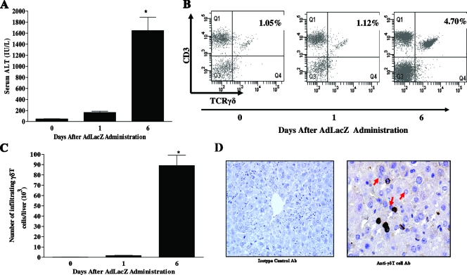
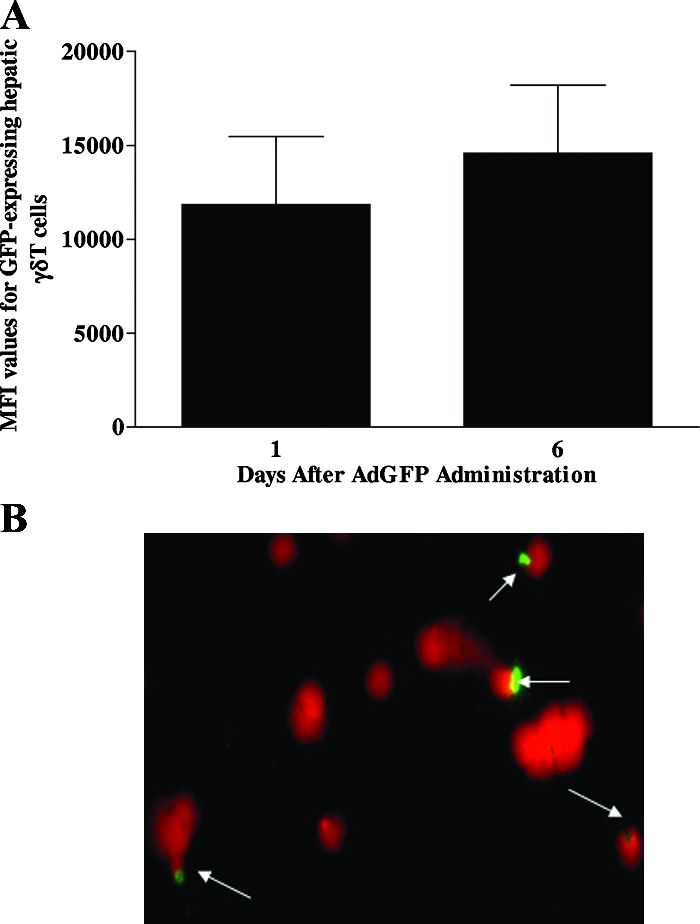
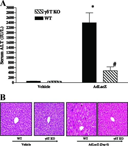
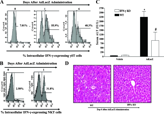
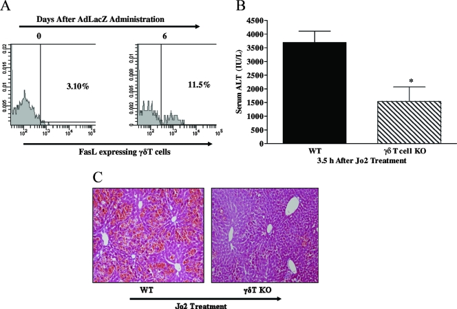
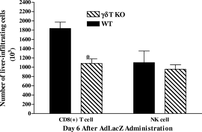
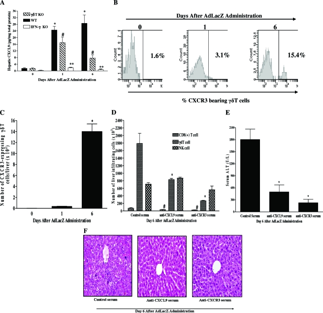
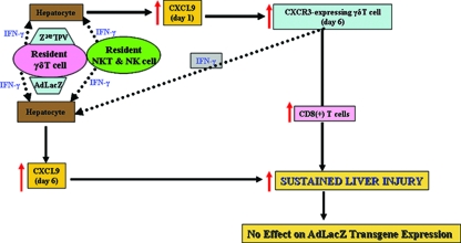
Emerging studies suggest an important role for the innate immune response in replication-defective adenovirus (Ad)-mediated acute liver toxicity. Specifically, classical innate immune cells (including NK cells, neutrophils, and Kupffer cells) have all been implicated in the development of Ad-mediated acute liver toxicity. The nonclassical innate immune T cell, the gammadeltaT cell, has been implicated in the pathophysiology of several viral infections that predominantly affect the mucosa and brain, but the specific role in the pathology of AdLacZ-mediated acute liver inflammation and injury as well as accompanying vector clearance is largely unknown. In the present study, we demonstrated that a CXCL9-CXCR3-dependent mechanism governed the accumulation of gammadeltaT cells in the livers of mice infected with Ad expressing the Escherichia coli LacZ gene (AdLacZ). We also showed a critical role for gammadeltaT cells in initiating acute liver toxicity after AdLacZ administration, driven in part by the ability of gammadeltaT cells to promote the recruitment of the conventional T cell, the CD8(+) T cell, into the liver. Furthermore, reduced hepatic injury in AdLacZ-infected gammadeltaT-cell-deficient mice was associated with lower hepatic levels of gamma interferon (IFN-gamma) and CXCL9, an IFN-gamma-inducible chemokine. Finally, our study highlighted a key role for IFN-gamma and CXCL9 cross talk acting in a feedback loop to drive the proinflammatory effects of gammadeltaT cells during AdLacZ-mediated acute liver toxicity. Specifically, intracellular IFN-gamma produced by activated hepatic gammadeltaT cells interacts with hepatocytes to mediate hepatic CXCL9 production, with the consequent accumulation of CXCR3-bearing gammadeltaT cells in the liver to cause acute liver damage without vector clearance.


Figures

FIG. 1.
FIG. 1.
Kinetics of acute liver damage and hepatic γδT-cell accumulation following AdLacZ administration. (A) C57BL/6 mice were given vehicle or infected with AdLacZ. Serum was collected at the indicated times and used to measure ALT levels (as described in Materials and Methods). Data are shown as means ± SEM (n = 5 to 8 mice per group; *, P ≤ 0.05 versus uninfected group, i.e., day 0). (B and C) To determine γδT-cell accumulation in the livers of C57BL/6 mice systemically challenged with AdLacZ or vehicle, hepatic lymphoid cells were isolated at the indicated times, stained with specific fluorochrome-labeled TCRγδ and CD3 MAbs, and then analyzed by flow cytometry to reveal the percentages (B) and absolute numbers (C) of γδT cells (i.e., TCRγδ-CD3 double-positive T cells) per liver. Data are presented as means ± SEM (n = 5 or 6 mice per group; *, P ≤ 0.05 versus uninfected group, i.e., day 0). (D) To reveal the localization of γδT cells in the liver, frozen liver sections were obtained from AdLacZ-infected C57BL/6 mice 6 days after infectivity and immunohistochemical localization of γδT cells was performed. Liver sections were stained with isotype control Ab or anti-γδT-cell Ab. γδT cells (white arrows) and hepatocytes (red arrows) are shown. Original magnification, ×400.
FIG. 2.
FIG. 2.
Ad interacts with γδT cells. (A) Male C57BL/6 mice were injected with AdGFP, and isolated hepatic fluorochrome-labeled γδT cells were screened for the presence of GFP by flow cytometry. The median fluorescence intensity (MFI) values for GFP-positive hepatic γδT cells are shown. Data are reported as means ± SEM (n = 4 mice per group). (B) To determine if Ad could interact with γδT cells, splenocytes were isolated from naive C57BL/6 mice and γδT cells were purified using a mouse γδT-cell isolation kit. Purified splenic γδT cells were then incubated in vitro with GFP-labeled Ad (104 virus particles) for 60 min. A fluorescence microscopic technique was used to evaluate Ad interaction with splenic γδT cells. Ad-GFP is shown in green, whereas γδT-cell nuclei counterstained with propidium iodide are shown in red.
FIG. 3.
FIG. 3.
Effects of γδT-cell deficiency on acute liver inflammation and injury in AdLacZ-infected mice. WT and γδT-cell KO mice were infected with AdLacZ or vehicle for 6 days. (A) Serum samples were obtained for the determination of ALT levels. All results are presented as means ± SEM (n = 6 mice per group; *, P ≤ 0.05 versus all vehicle-treated controls; #, P ≤ 0.05 versus AdLacZ-infected WT mice). (B) Photomicrograph of H&E-stained representative liver sections depicting diffuse and severe acute hepatic injury with swelling of the hepatocytes, obliteration of the sinusoid spaces, hepatocellular necrosis, and numerous acidophilic bodies in AdLacZ-infected WT mice compared to the minimal distortion of lobular architecture and absence of inflammatory cell infiltrates in liver sections from γδT-cell KO mice after 6 days of AdLacZ infection. Original magnification, ×200. In contrast, H&E-stained liver sections from vehicle-treated WT and γδT-cell KO mice were normal.
FIG. 4.
FIG. 4.
Role of IFN-γ in the development of acute liver inflammation and injury in mice systemically challenged with AdLacZ. (A) To determine if γδT cells are a source of hepatic IFN-γ after AdLacZ infectivity, C57BL/6 mice were systemically challenged with AdLacZ or vehicle and hepatic lymphoid cells were isolated at the indicated times. Isolated hepatic lymphoid cells were first stained with specific fluorochrome-labeled TCRγδ and CD3 MAbs to identify γδT cells, and the cells were then permeabilized prior to staining with fluorochrome-labeled IFN-γ MAb to determine γδT-cell intracellular IFN-γ after flow cytometric analysis. A representative FACS histogram demonstrating intracellular IFN-γ expression by isolated hepatic γδT cells after AdLacZ administration is shown. (B) Next, we determined if hepatic NKT cells are also an early source of intracellular IFN-γ after AdLacZ administration. C57BL/6 mice were systemically challenged with AdLacZ or vehicle, and hepatic lymphoid cells were isolated 24 h later. Isolated hepatic lymphoid cells were first stained with specific fluorochrome-labeled TCRβ MAb and the NKT cell tetramer CD1d-PBS57 to identify NKT cells (see Materials and Methods), and the cells were then permeabilized prior to staining with fluorochrome-labeled IFN-γ MAb. A representative FACS histogram demonstrating intracellular IFN-γ expression by isolated hepatic NKT cells after AdLacZ or vehicle administration at day 1 posttreatment is shown. (C) To determine the specific contribution of IFN-γ to acute liver injury mediated by systemically administered AdLacZ, WT and IFN-γ KO mice were infected with AdLacZ for 6 days. Serum samples were obtained for the determination of ALT levels. All results are presented as means ± SEM (n = 5 or 6 mice per group; *, P ≤ 0.05 versus all vehicle-treated controls; #, P ≤ 0.05 versus AdLacZ-infected WT mice). (D) Photomicrograph of H&E-stained representative liver sections depicting diffuse and severe acute hepatic injury (inflammation and hepatocellular necrosis with numerous acidophilic bodies) in AdLacZ-infected WT mice compared with essentially normal liver histology in AdLacZ-infected IFN-γ KO mice. Original magnification, ×200.
FIG. 5.
FIG. 5.
Role of the Fas/FasL-mediated death pathway in the proinflammatory effect of γδT cells. (A and B) Agonistic anti-Fas antibody was used to specifically determine if γδT cells are capable of promoting acute liver damage via the Fas-mediated death pathway. WT and γδT-cell KO mice were administered the agonistic anti-Fas antibody Jo2 (0.5 μg/g body weight; intraperitoneally), and acute liver injury (i.e., ALT levels and histology) was assessed 3.5 h later. Data are presented as means ± SEM (n = 4 to 7 mice per group; *, P ≤ 0.05 versus WT mice). Photomicrograph of H&E-stained representative liver sections depicting diffuse and severe acute liver failure (inflammation and widespread hepatocellular necrosis with numerous apoptosis) in Jo2-treated WT mice compared with patchy necrosis and reduced inflammation in a liver sections from γδT-cell KO mice administered Jo2. Original magnification, ×200. (C) C57BL/6 mice were administered vehicle or AdLacZ, and hepatic lymphoid cells were isolated from these mice 6 days after infectivity. Isolated hepatic lymphoid cells were initially stained with fluorochrome-labeled TCRγδ and CD3 MAbs to identify γδT cells and then stained with fluorochrome-labeled FasL MAb to determine FasL cell surface expression on hepatic γδT cells. A representative FACS histogram demonstrating extracellular FasL expression on γδT cells after AdLacZ administration is depicted.
FIG. 6.
FIG. 6.
Effects of γδT-cell deficiency on NK cell and CD8+ T-cell accumulation in the livers of AdLacZ-infected mice. WT and γδT-cell KO mice were infected with AdLacZ, and hepatic lymphoid cells were isolated 6 days later. Isolated hepatic lymphoid cells were then stained with fluorochrome-labeled NK1.1 MAb and fluorochrome-labeled CD8a MAb and analyzed by flow cytometry to identify accumulation of hepatic NK and CD8+ T cells, respectively. The absolute numbers of NK cells and CD8+ T cells are shown. All results are presented as means ± SEM (n = 5 or 6 mice per group; *, P ≤ 0.05 versus AdLacZ-infected WT mice).
FIG. 7.
FIG. 7.
Role of the CXCR3-CXCL9 pathway in the effector function of γδT cells during AdLacZ-induced liver inflammation and injury. (A) Kinetics of hepatic CXCL9 production during AdLacZ-mediated liver toxicity was evaluated using a Bioplex array system (see Materials and Methods). The effects of γδT-cell deficiency and IFN-γ deficiency on hepatic CXCL9 production after AdLacZ administration were also evaluated. Results are presented as means ± SEM (n = 5 or 6 mice per group; *, P ≤ 0.05 versus uninfected group, i.e., day 0; #, P ≤ 0.05 versus AdLacZ-infected WT mice; **, P ≤ 0.05 versus AdLacZ-infected WT mice). (B and C) Flow cytometric analysis was used to determine the kinetics of CXCR3 expression on hepatic γδT cells during AdLacZ infection. C57BL/6 mice were uninfected (day 0) or infected with AdLacZ, and hepatic lymphoid cells were isolated at the indicated times. Isolated cells were then stained with specific fluorochrome-labeled TCRγδ, CD3, and CXCR3 MAbs to reveal the percentage (B) and absolute number (C) of hepatic γδT cells expressing CXCR3 receptors. Data are presented as means ± SEM (n = 5 or 6 mice per group; *, P ≤ 0.05 versus uninfected group, i.e., day 0). (D) To determine the functional role of CXCR3 and CXCL9 in the recruitment of γδT cells, NK cells, and CD8+ T cells to the liver following AdLacZ administration, anti-CXCR3 serum and anti-CXCL9 serum were used. C57BL/6 mice were given control serum, anti-CXCR3 serum, or anti-CXCL9 serum intraperitoneally 16 h before AdLacZ infection, with additional doses given every 48 h until termination of the experiment. All mice were sacrificed at day 6 after AdLacZ administration for the determination of hepatic γδT-cell, NK cell, and CD8+ T-cell accumulation by flow cytometry. Data are presented as means ± SEM (n = 5 or 6 mice per group; *, P ≤ 0.05 versus control serum; #, P ≤ 0.05 versus control serum). (E and F) The effects of anti-CXCR3 serum and anti-CXCL9 serum treatment on liver injury 6 days after AdLacZ infection were also determined biochemically by measuring ALT levels (E) and by histology (F). ALT values are depicted as means ± SEM (n = 6 to 8 mice per group; *, P ≤ 0.05 versus control serum). Panel F shows a representative photomicrograph of liver section from AdLacZ-infected mice treated with control serum, which was characterized by diffuse and severe acute hepatic injury, whereas liver sections from anti-CXCR3 serum-treated and anti-CXCL9-treated mice were characterized by nearly normal liver histology with no hepatocellular injury following the administration of AdLacZ. Original magnification, ×200.
FIG. 8.
FIG. 8.
Schematic overview of the potential mechanisms that promote the proinflammatory effect of γδT cells during AdLacZ-mediated acute liver toxicity (see Discussion).

Similar articles

Cited by

References

    1. Ajuebor, M. N., A. I. Aspinall, F. Zhou, T. Le, Y. Yang, S. J. Urbanski, S. Sidobre, M. Kronenberg, C. M. Hogaboam, and M. G. Swain. 2005. Lack of chemokine receptor CCR5 promotes murine fulminant liver failure by preventing the apoptosis of activated CD1d-restricted NKT cells. J. Immunol. 1748027-8037. - PubMed
    1. Ajuebor, M. N., C. M. Hogaboam, T. Le, A. E. Proudfoot, and M. G. Swain. 2004. CCL3/MIP-1α is pro-inflammatory in murine T cell-mediated hepatitis by recruiting CCR1-expressing CD4+ T cells to the liver. Eur. J. Immunol. 342907. - PubMed
    1. Ajuebor, M. N., C. M. Hogaboam, T. Le, and M. G. Swain. 2003. C-C chemokine ligand 2/monocyte chemoattractant protein-1 directly inhibits NKT cell IL-4 production and is hepatoprotective in T cell-mediated hepatitis in the mouse. J. Immunol. 1705252-5259. - PubMed
    1. Ajuebor, M. N., and M. G. Swain. 2002. Role of chemokines and chemokine receptors in the gastrointestinal tract. Immunology 105137-143. - PMC - PubMed
    1. Ajuebor, M. N., M. G. Swain, and M. Perretti. 2002. Chemokines as novel therapeutic targets in inflammatory diseases. Biochem. Pharmacol. 631191-1196. - PubMed

Publication types