. 2008 Oct;19(10):4122-9.
doi: 10.1091/mbc.e07-10-1079. Epub 2008 Jul 30.

Microtubules have opposite orientation in axons and dendrites of Drosophila neurons

Affiliations

Microtubules have opposite orientation in axons and dendrites of Drosophila neurons

Michelle C Stone et al. Mol Biol Cell. 2008 Oct.

Abstract

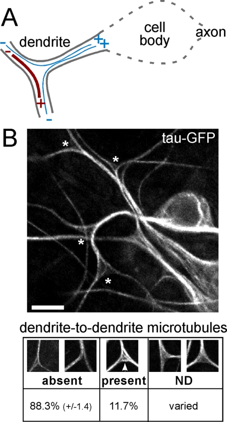
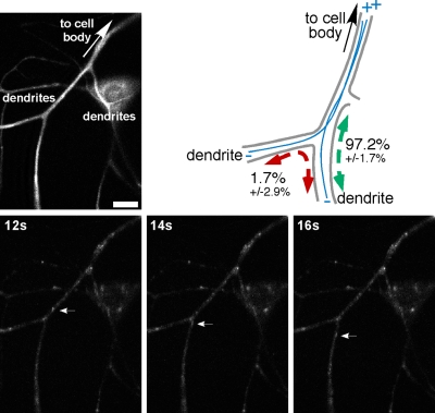
In vertebrate neurons, axons have a uniform arrangement of microtubules with plus ends distal to the cell body (plus-end-out), and dendrites have equal numbers of plus- and minus-end-out microtubules. To determine whether microtubule orientation is a conserved feature of axons and dendrites, we analyzed microtubule orientation in invertebrate neurons. Using microtubule plus end dynamics, we mapped microtubule orientation in Drosophila sensory neurons, interneurons, and motor neurons. As expected, all axonal microtubules have plus-end-out orientation. However, in proximal dendrites of all classes of neuron, approximately 90% of dendritic microtubules were oriented with minus ends distal to the cell body. This result suggests that minus-end-out, rather than mixed orientation, microtubules are the signature of the dendritic microtubule cytoskeleton. Surprisingly, our map of microtubule orientation predicts that there are no tracks for direct cargo transport between the cell body and dendrites in unipolar neurons. We confirm this prediction, and validate the completeness of our map, by imaging endosome movements in motor neurons. As predicted by our map, endosomes travel smoothly between the cell body and axon, but they cannot move directly between the cell body and dendrites.


Figures

Figure 1.
Figure 1.
Known microtubule orientation in vertebrate and Drosophila da neurons, and possible scenarios for the arrangement of microtubules in fly dendrites. (A) In frog mitral cells and cultured rodent interneurons, microtubules in dendrites have mixed orientation, whereas in Drosophila da neurons ∼95% have minus ends distal to the cell body based on EB1-GFP dynamics. In all neurons examined, plus-end-out microtubules predominate in axons. (B) The failure to find a significant population of plus-end-out microtubules in da neuron dendrites can be accounted for by several explanations. 1) The arrangement of microtubules in sensory da dendrites could be different from the arrangement in interneuron dendrites. 2) Analysis of microtubule orientation by EB1-GFP dynamics could have missed a significant population of stable plus-end-out microtubules. 3) Minus-end-out microtubules could predominate in all Drosophila neurons.
Figure 2.
Figure 2.
Microtubule orientation in dendrites of a type I da sensory neuron. (A) An overview of EB1-GFP in the region of the dorsal cluster of the peripheral nervous system in a live larva is shown. The cell body of ddaE is indicated with an arrow. Cell bodies of other peripheral neurons are to the left. The boxed region of ddaE dendrites was chosen for higher power imaging, and frames were acquired every 2 s. Three frames are shown in B. Two EB1-GFP dots that move toward the cell body are indicated with a white arrow and arrowhead, and a dot in a distal dendrite that moves away from the cell body is indicated with a gray arrow. (C) Microtubule orientation derived from this type of data is summarized in the table. Data were derived from four groups of larvae (3 groups of 9 larvae and 1 group of 10 larvae). A single da neuron was analyzed in each larva. In total, 143 EB1-GFP dots were counted in the main dendrite trunk and 54 dots in distal dendrites. (D) Diagram of microtubule orientation in da neurons. Bar, 20 μm (A) and 10 μm (B).
Figure 3.
Figure 3.
Microtubule orientation in an interneuron. (A) Overview of the single neuron in larval brains that expresses bright EB1-GFP in response to the RN2-Gal4. The image is a projection of a confocal stack. Dendrites branch from the primary neurite on the same side of the brain as the cell body. An axon crosses to the other brain lobe and makes terminal branches with large synaptic boutons. Approximate outline of brain lobes is indicated with dotted line. Image was rotated from original and placed on a black background. (B) Three frames from a movie of a different brain are shown. The area shown in the frames is similar to the boxed area in A, with the dendrite at the left. Two EB1-GFP dots, indicated by arrows, move toward the point at which the dendrite branches from the primary neurite, and thus toward the cell body. (C) Quantitation of EB1-GFP movements. Dots were tracked in the axon just beyond the point at which the dendrites branch off, in proximal dendrites, and in the primary neurite. Two groups of six and one group of five brains were analyzed, with a total of 64 EB1-GFP dots. Bar, 25 μm (A) and 10 μm (B).
Figure 4.
Figure 4.
Microtubule orientation in motor neurons. (A) Three consecutive frames from a movie of EB1-GFP dynamics in the ventral ganglion of a brain explant are shown. EB1-GFP expression is controlled by RN2-Gal4. The motor neuron axons were easily identifiable as they leave the ventral ganglion in a motor nerve. Dendrites branch from the primary neurite and make complex arborizations in the ventral ganglion. We focused mostly on the proximal region of the dendrite to avoid ambiguities from crossing dendrite branches. Arrows indicate an EB1-GFP dot in a dendrite that moves toward the cell body and thus is at the tip of a minus-end-out microtubule. (B) Quantitation of microtubule orientation in motor neuron axons and dendrites derived from EB1-GFP dynamics. We were unable to quantitate data in the primary neurite due to overlapping structures. Axon data were acquired from two groups of six and one group of seven brains, with a total of 45 EB1-GFP dots counted. Dendrite data were acquired from two sets of 12 and one set of 13 brains, with a total of 69 dots counted. (C) Diagram of microtubule orientation in unipolar neurons, based on data from the interneuron in Figure 3 and motor neurons. Bar, 5 μm (A).
Figure 5.
Figure 5.
The arrangement of stable microtubules at da dendrite branch points is consistent with most microtubules in dendrites having the same orientation. (A) EB1-GFP dynamics indicated that minus-end-out microtubules are present in da dendrites (thin blue lines). To determine whether plus-end-out microtubules are also abundant, we assayed dendrite branch points for microtubules that form dendrite-to-dendrite bridges (thick red line). (B) A tau-GFP protein trap line was used to visualize microtubules in da dendrites in whole, living larvae. A single plane from an image stack acquired with a confocal microscope is shown. Stars indicate triangular branch points with clear microtubules that run between the cell body and dendrite branches, but not from one dendrite branch to another. Examples of different classes of branch points are shown: first two images are examples of branches in which a dendrite-to-dendrite microtubule is absent, third image is a branch in which a dendrite-to-dendrite microtubule is present (arrow points to dendrite-to-dendrite microtubule), and last two images were not determined (ND) because the image was not clear at the branch. In each panel, the top of the image is closest to the cell body. Branches were classified into these groups in three experiments with slightly different imaging conditions. The number of ND branches varied depending on the imaging conditions, and so was not included in the percentage. The percentage of branches without dendrite-to-dendrite microtubules varied very little. We analyzed 117 images, with 194 branches in the first two categories, and 88 ND. Bar, 5 μm (B).
Figure 6.
Figure 6.
Routes of endosome transport in da neuron dendrites. Rab4-RFP was imaged in da neurons in larvae heterozygous for the 109(2)80 Gal4 driver, UAS-EB1-GFP, and UAS-Rab4-RFP. Movies were acquired at 2-s intervals. For the example shown, the first image from the EB1-GFP track shows the layout of the dendrites (top left). The cell body is out of the frame at the top. These neurons are multipolar, so all processes in the image are dendrites. Three frames from the Rab4-RFP track are shown. An endosome moving from the cell body side of the branch out into a distal dendrite is indicated with an arrow. Movements of endosomes through dendrite branch points are summarized in the diagram. Predicted microtubule tracks are drawn in blue. Endosomes that moved through a branch were categorized as traveling between cell body and distal dendrite (green double headed arrow), or from dendrite to dendrite (red double arrow). Two groups of six larvae and one group of five were analyzed, and the total number of endosomes counted was 43. Bar, 5 μm.
Figure 7.
Figure 7.
Routes of endosome transport in motor neurons. Endosomes in motor neurons were labeled with RFP in flies homozygous for RN2-Gal4, UAS-EB1-GFP, and UAS-Rab4-RFP. Movies were acquired at 1- or 2-s intervals: 1 s in the example shown. Two frames in different focal planes from the EB1-GFP track were summed to give an overview (top left). The cell body is out of the frame at the top, and the axon exits at the bottom. Five frames from the Rab4-RFP track are shown. An endosome that travels from the dendrites to the axon is indicated with arrows. The whole movie is available as Supplemental Movie 5. A summary of routes taken through the primary neurite–axon–dendrite branch is shown at top right. The predicted layout of microtubules is indicated by blue lines. Endosomes that moved through the branch were classified as moving between the cell body and dendrites (red double arrow), between cell body and axon (green double arrow at right), or between dendrite and axon (green double arrow at left). Two groups of five, and one group of six, larval brains were analyzed. Total number of endosomes counted was 34. Bar, 5 μm.

Similar articles

Cited by

References

    1. Alberts B., Johnson A., Lewis J., Raff M., Roberts K., Walter P. Molecular Biology of the Cell. New York: Garland Science; 2002.
    1. Baas P. W., Deitch J. S., Black M. M., Banker G. A. Polarity orientation of microtubules in hippocampal neurons: uniformity in the axon and nonuniformity in the dendrite. Proc. Natl. Acad. Sci. USA. 1988;85:8335–8339. - PMC - PubMed
    1. Baas P. W., White L. A., Heidemann S. R. Microtubule polarity reversal accompanies regrowth of amputated neurites. Proc. Natl. Acad. Sci. USA. 1987;84:5272–5276. - PMC - PubMed
    1. Black M. M., Baas P. W. The basis of polarity in neurons. Trends Neurosci. 1989;12:211–214. - PubMed
    1. Burack M. A., Silverman M. A., Banker G. The role of selective transport in neuronal protein sorting. Neuron. 2000;26:465–472. - PubMed

Publication types

Substances

LinkOut - more resources