Comparative Study
. 2008 Jul 30;28(31):7774-80.
doi: 10.1523/JNEUROSCI.1069-08.2008.

Facilitation of stepping with epidural stimulation in spinal rats: role of sensory input

Affiliations
Comparative Study

Facilitation of stepping with epidural stimulation in spinal rats: role of sensory input

Igor Lavrov et al. J Neurosci. .

Abstract

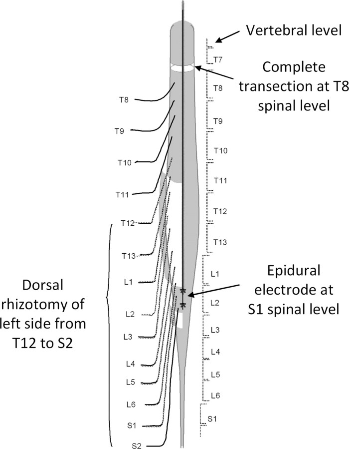
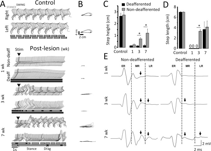
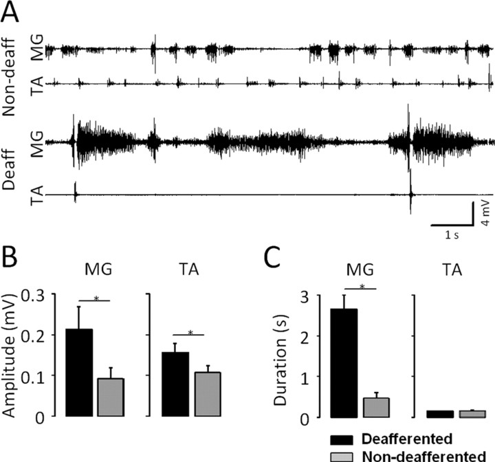
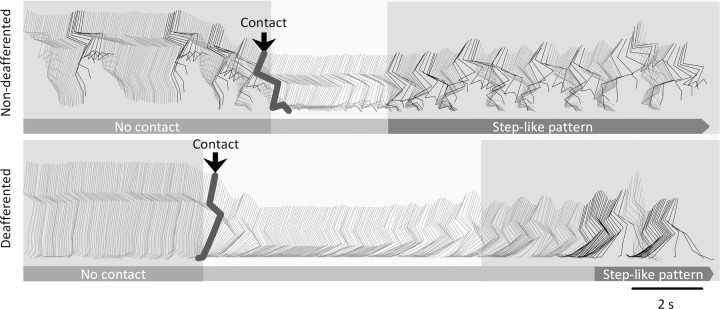
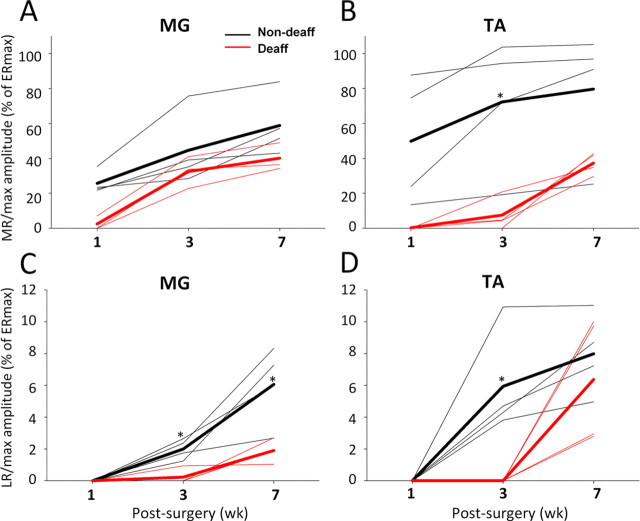
We investigated the role of afferent information during recovery of coordinated rhythmic activity of the hindlimbs in rats with a complete spinal cord section (approximately T8) and unilateral deafferentation (T12-S2) to answer the following questions: (1) Can bilateral stepping be generated with only afferent projections intact on one side? (2) Can the sensory input from the non-deafferented side compensate for the loss of the afferent input from the deafferented side through the crossed connections within the lumbosacral spinal cord? (3) Which afferent projections to the spinal cord from the non-deafferented side predominantly mediate the effect of epidural stimulation to facilitate stepping? Recovery of stepping ability was tested under the facilitating influence of epidural stimulation at the S1 spinal segment, or epidural stimulation plus quipazine, a 5-HT agonist. All chronic spinal rats were able to generate stepping-like patterns on a moving treadmill on the non-deafferented, but not deafferented, side from 3 to 7 weeks after surgery when facilitated by epidural stimulation. Adaptation to the loss of unilateral afferent input was evident at 7 weeks after surgery, when some movements occurred on the deafferented side. Spinal-cord-evoked potentials were observed on both sides, although middle (monosynaptic) and late (long latency) responses were more prominent on the non-deafferented side. The afferent information arising from the non-deafferented side, however, eventually could mediate limited restoration of hindlimb movements on the deafferented side. These data suggest that facilitation of stepping with epidural stimulation is mediated primarily through ipsilateral afferents that project to the locomotor networks.


Conflict of interest statement

The authors declare no competing financial interests.

Figures

Figure 1.
Figure 1.
Schematic of the spinal cord transection and unilateral deafferentation surgery. A complete spinal cord transection was performed at approximately the T8 spinal level (between vertebral segments T7 and T8). The dura (gray shading) was opened longitudinally from vertebral level T11 to S3 (leaving a short strip at approximately the L2 level intact), and a unilateral (left side) dorsal rhizotomy (cutting the dorsal root as close to the spinal cord as possible and as close to its exit from the vertebral column as possible) was performed intradurally from the T12 to the S2 spinal segments. A single insulated wire electrode with a small notch (∼0.5–1.0 mm) exposing the wire facing the spinal cord was sutured (asterisks represent the two sutures) on the short strip of intact dura over spinal segment S1 through which electrical stimulation was used to facilitate stepping and induce bilateral reflex responses. The middle arrow points to the transection site, and the bottom arrow, to the epidural electrode implant site. The extent of the unilateral dorsal rhizotomy is identified on the left side with the cut dorsal roots shown as broken lines.
Figure 2.
Figure 2.
The rhythmicity of activity facilitated by epidural stimulation (40 Hz) and of spinal cord reflexes to a single stimulus after a complete midthoracic spinal cord transection and unilateral dorsal rhizotomy (Fig. 1). A, Representative stick diagrams of the hindlimb movements in a control rat and in an experimental rat, 1, 3, and 7 weeks after lesion. The time between individual sticks is 40 ms. Blocks at the bottom of each pair of stick diagrams represent stance (gray), swing (blank), and foot dragging (black). Arrowheads indicate the initiation of epidural stimulation (Stim). Deaff, Deafferentation; Non-deaff, non-deafferentation. B, Successive trajectories of the right and left hindlimb endpoint recorded from the same rats and same steps as in A. C, D, Mean (SEM) step height (C) and step length (D) from both limbs of control and spinal-transected rats with unilateral deafferention. E, Spinal cord motor-evoked responses to a single stimulus recorded from the tibialis anterior muscle on the non-deafferented and deafferented hindlimb of a representative spinal rat at 1, 3, and 7 weeks after lesion. The stimulus (not shown) is at the beginning of each trace. The arrows indicate the appearance of MR or LR. *p < 0.05, Significant difference between the non-deafferented and deafferented hindlimbs. All values for the non-deafferented and deafferented hindlimbs in C and D are statistically lower than control. ER, Early response. Error bars indicate SEM.
Figure 3.
Figure 3.
Analysis of EMG activity on the non-deafferented (Non-deaff) and deafferented (Deaff) sides at 7 weeks after a complete spinal cord transection and unilateral deafferentation. A, EMG bursting patterns for the MG and TA muscles on the non-deafferented and deafferented hindlimbs of a representative spinal rat during treadmill stepping at 7 cm/s facilitated by epidural stimulation (at S1, 40 Hz) and quipazine (0.3 mg/kg, i.p.) administration. B, C, Mean (SEM) EMG burst amplitude (B) and duration (C) for the MG and TA in the non-deafferented and deafferented hindlimbs for all rats. *p < 0.05, significant difference between the non-deafferented and deafferented hindlimbs. Error bars indicate SEM.
Figure 4.
Figure 4.
Intact afferents are required for the initiation of activity on the non-deafferented and deafferented sides in spinal rats. Representative stick diagrams are shown of the movements observed on the non-deaffererented and deafferented sides of a spinal rat while suspended above the treadmill (no foot contact) and after contact (arrows) of the feet with the moving treadmill belt when facilitated by epidural stimulation (at S1, 40 Hz) and quipazine (0.3 mg/kg, i.p.) administration. The time between individual sticks is 40 ms. Note the slow and delayed movements on the deafferented side occurring after rhythmic activity is initiated on the non-deafferented side.
Figure 5.
Figure 5.
A–D, Changes in the maximum amplitude of the MR (MR/max) and LR (LR/max) for the MG (A, C) and TA (B, D) on the non-deafferented (black) and deafferented (red) sides of a spinal rat at 1, 3, and 7 weeks after surgery. Data represent the average of 10 evoked potentials from four rats (thin lines) and the overall average for the four rats for each time point (thick lines). MR/max and LR/max are shown as a percentage of ERmax at each time point. Note that the amplitudes of the responses are consistently higher on the non-deafferented side and that a significant level of recovery of the LR on the deafferented side occurs only at 7 weeks for both the MG and TA. *p < 0.05, significant difference between the means for the non-deafferented and deafferented hindlimbs.

References

    1. Coburn B. A theoretical study of epidural electrical stimulation of the spinal cord–Part II: Effects on long myelinated fibers. IEEE Trans Biomed Eng. 1985;32:978–986. - PubMed
    1. Coburn B, Sin WK. A theoretical study of epidural electrical stimulation of the spinal cord–Part I: Finite element analysis of stimulus fields. IEEE Trans Biomed Eng. 1985;32:971–977. - PubMed
    1. Courtine G, Roy RR, Hodgson J, McKay H, Raven J, Zhong H, Yang H, Tuszynski MH, Edgerton VR. Kinematic and EMG determinants in quadrupedal locomotion of a non-human primate (Rhesus) J Neurophysiol. 2005;93:3127–3145. - PubMed
    1. Courtine G, Harkema SJ, Dy CJ, Gerasimenko YP, Dyhre-Poulsen P. Modulation of multisegmental monosynaptic responses in a variety of leg muscles during walking and running in humans. J Physiol. 2007;582:1125–1139. - PMC - PubMed
    1. Courtine G, Song B, Roy RR, Zhong H, Herrmann JE, Ao Y, Qi J, Edgerton VR, Sofroniew MV. Recovery of supraspinal control of stepping via indirect propriospinal relay connections after spinal cord injury. Nat Med. 2008;14:69–74. - PMC - PubMed

Publication types