Functional convergence of dopaminergic and cholinergic input is critical for hippocampus-dependent working memory
- PMID: 18667612
- PMCID: PMC6670368
- DOI: 10.1523/JNEUROSCI.1885-08.2008
Functional convergence of dopaminergic and cholinergic input is critical for hippocampus-dependent working memory
Abstract
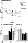
Although Parkinson's disease is a movement disorder, in many patients cognitive dysfunction is an important clinical sign. It is not yet clear whether this is attributable solely to a decrease in dopamine levels, or whether other neurotransmitter systems might be involved as well. In the present study, the importance of the mesocorticolimbic dopamine pathway and a possible convergence with forebrain cholinergic projections to neocortex and hippocampus in the regulation of learning and memory abilities were investigated by using specific lesion paradigms in one or both systems. Lesioning of dopaminergic neurons in the ventral tegmental area resulted in an impaired performance in the reference memory task, whereas the execution of the working memory tasks appeared to be unaffected in the Morris water maze. Analysis of the swim paths revealed that the dopamine-depleted animals were capable of adapting a search strategy on a given testing day but failed to transfer this information to the next day, suggesting a deficit in information storage and/or recall. In contrast, cholinergic lesions alone were without effect in all test paradigms. However, when both dopamine and acetylcholine were depleted, animals were also impaired in the working memory task, indicating that a functional convergence of the inputs from these systems was critical for acquisition of spatial memory. Interestingly, such an additional acquisition deficit appeared only after hippocampal cholinergic depletion regardless of a concurrent disruption of basalo cortical cholinergic afferents. Thus, further analyses of cholinergic alterations may prove useful in better understanding the cognitive symptoms in Parkinson's disease.
Figures


Similar articles
-
Amelioration of spatial navigation and short-term memory deficits by grafts of foetal basal forebrain tissue placed into the hippocampus and cortex of rats with selective cholinergic lesions.Eur J Neurosci. 1998 Jul;10(7):2353-70. doi: 10.1046/j.1460-9568.1998.00247.x. Eur J Neurosci. 1998. PMID: 9749764
-
Spatial learning and memory following fimbria-fornix transection and grafting of fetal septal neurons to the hippocampus.Exp Brain Res. 1987;67(1):195-215. doi: 10.1007/BF00269466. Exp Brain Res. 1987. PMID: 3622677
-
Effects of cholinergic-rich neural grafts on radial maze performance of rats after excitotoxic lesions of the forebrain cholinergic projection system--I. Amelioration of cognitive deficits by transplants into cortex and hippocampus but not into basal forebrain.Neuroscience. 1991;45(3):587-607. doi: 10.1016/0306-4522(91)90273-q. Neuroscience. 1991. PMID: 1775235 Review.
-
Cholinergic system and memory in the rat: effects of chronic ethanol, embryonic basal forebrain brain transplants and excitotoxic lesions of cholinergic basal forebrain projection system.Neuroscience. 1989;33(3):435-62. doi: 10.1016/0306-4522(89)90397-7. Neuroscience. 1989. PMID: 2636702
-
Storage, recall, and novelty detection of sequences by the hippocampus: elaborating on the SOCRATIC model to account for normal and aberrant effects of dopamine.Hippocampus. 2001;11(5):551-68. doi: 10.1002/hipo.1071. Hippocampus. 2001. PMID: 11732708 Review.
Cited by
-
Amelioration of the haloperidol-induced memory impairment and brain oxidative stress by cinnarizine.EXCLI J. 2012 Aug 27;11:517-30. eCollection 2012. EXCLI J. 2012. PMID: 27540345 Free PMC article.
-
Modelling the dopamine and noradrenergic cell loss that occurs in Parkinson's disease and the impact on hippocampal neurogenesis.Hippocampus. 2018 May;28(5):327-337. doi: 10.1002/hipo.22835. Epub 2018 Feb 24. Hippocampus. 2018. PMID: 29431270 Free PMC article.
-
Ventral tegmental area disruption selectively affects CA1/CA2 but not CA3 place fields during a differential reward working memory task.Hippocampus. 2011 Feb;21(2):172-84. doi: 10.1002/hipo.20734. Hippocampus. 2011. PMID: 20082295 Free PMC article.
-
Spatial Memory Performance Associated with Measures of Immune Function in Elderly Female Rhesus Macaques.Eur Geriatr Med. 2011 Apr 1;2(2):117-121. doi: 10.1016/j.eurger.2011.01.002. Eur Geriatr Med. 2011. PMID: 21603071 Free PMC article.
-
Idebenone improves motor dysfunction, learning and memory by regulating mitophagy in MPTP-treated mice.Cell Death Discov. 2022 Jan 17;8(1):28. doi: 10.1038/s41420-022-00826-8. Cell Death Discov. 2022. PMID: 35039479 Free PMC article.
References
-
- Aarsland D, Andersen K, Larsen JP, Lolk A, Kragh-Sørensen P. Prevalence and characteristics of dementia in Parkinson disease: an 8-year prospective study. Arch Neurol. 2003;60:387–392. - PubMed
-
- Agid YA, Javoy-Agid F, Ruberg M. Biochemistry of neurotransmitters in Parkinson's disease. In: Marsden CD, Fahn S, editors. Movement disorders 2. London: Butterworths; 1987. pp. 166–230.
-
- Bartus RT, Dean RL, 3rd, Beer B, Lippa AS. The cholinergic hypothesis of geriatric memory dysfunction. Science. 1982;217:408–414. - PubMed
-
- Bigl V, Woolf NJ, Butcher LL. Cholinergic projections from the basal forebrain to frontal, parietal, temporal, occipital, and cingulate cortices: a combined fluorescent tracer and acetylcholinesterase analysis. Brain Res Bull. 1982;8:727–749. - PubMed
Publication types
MeSH terms
Substances
LinkOut - more resources
Full Text Sources
Medical
