Comparative Study
. 2008 Jul 30;28(31):7919-28.
doi: 10.1523/JNEUROSCI.0784-08.2008.

Diverse mechanisms underlie glycinergic feedback transmission onto rod bipolar cells in rat retina

Affiliations
Comparative Study

Diverse mechanisms underlie glycinergic feedback transmission onto rod bipolar cells in rat retina

Andrés E Chávez et al. J Neurosci. .

Abstract

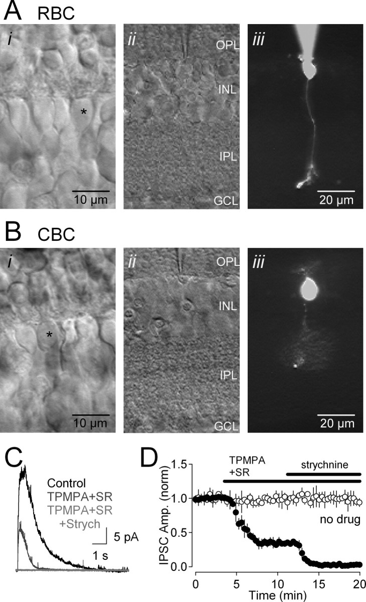
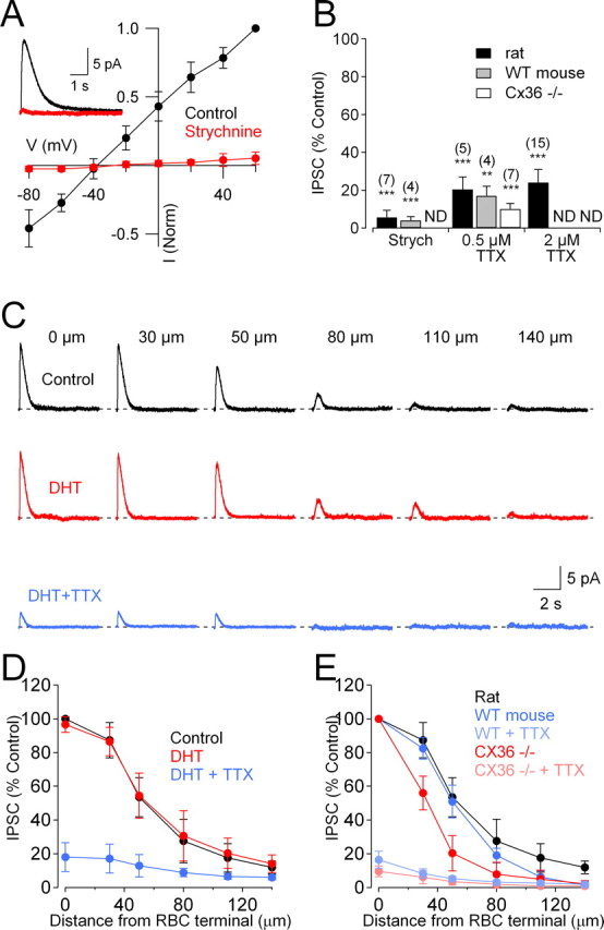
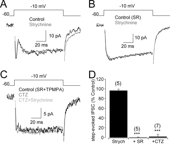
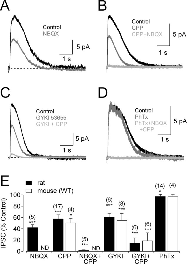
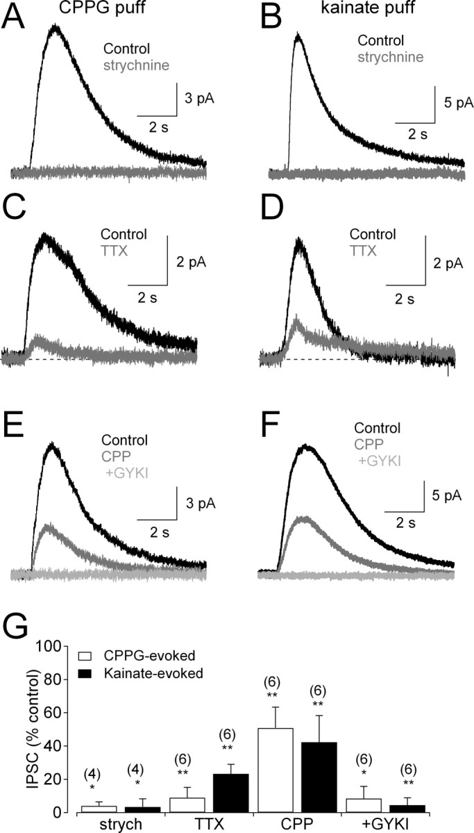
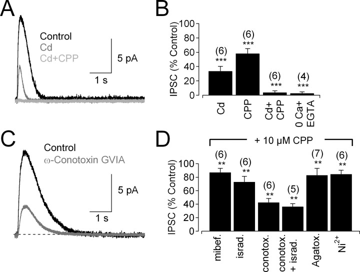
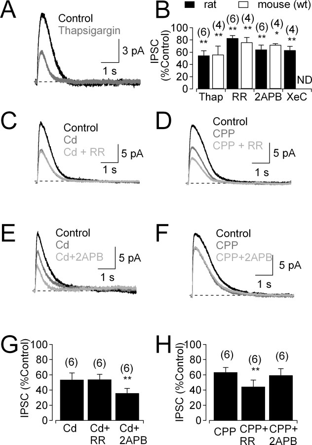
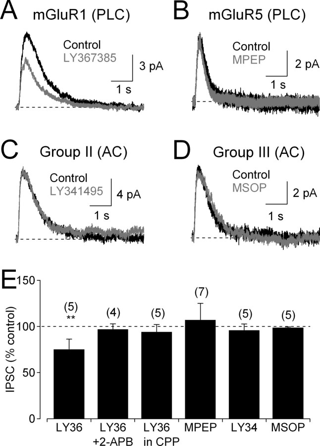
Synaptic inhibition shapes visual signaling in the inner retina, but the physiology of most amacrine cells, the interneurons that mediate this inhibition, is poorly understood. Discerning the function of most individual amacrine cell types is a daunting task, because few molecular or morphological markers specifically distinguish between approximately two dozen different amacrine cell types. Here, we examine a functional subset of amacrine cells by pharmacologically isolating glycinergic inhibition and evoking feedback IPSCs in a single cell type, the rod bipolar cell (RBC), with brief glutamate applications in the inner plexiform layer. We find that glycinergic amacrine cells innervating RBCs receive excitatory inputs from ON and OFF bipolar cells primarily via NMDA receptors (NMDARs) and Ca2+-impermeable AMPA-type glutamate receptors. Glycine release from amacrine cells is triggered by Ca2+ influx through both voltage-gated Ca2+ (Ca(v)) channels and NMDARs. These intracellular Ca2+signals are amplified by Ca2+-induced Ca2+ release via both ryanodine and IP3 receptors, which are activated independently by Ca2+ influx through Ca(v) channels and NMDARs, respectively. Glycinergic feedback signaling depends strongly, although not completely, on voltage-gated Na+ channels, and the spatial extent of feedback inhibition is expanded by gap junction connections between glycinergic amacrine cells. These results indicate that a diversity of mechanisms underlie glycinergic feedback inhibition onto RBCs, yet they highlight several physiological themes that appear to distinguish amacrine cell function.


Figures

Figure 1.
Figure 1.
Whole-cell recordings of inhibitory feedback in RBCs in retinal slices. A, Micrographs of an RBC in a retinal slice. i, High-magnification DIC image of the INL, showing bipolar cell somata. ii, iii, The selected cell (*) was patched (ii), filled with Alexa-488, and imaged under epifluorescence (iii). The scale bar in iii also applies to ii. B, As in A, but recording from a cone bipolar cell (CBC). C, Feedback IPSCs elicited in an RBC (Vhold = 0 mV) were blocked partially by GABAR antagonists TPMPA and SR95531 (SR); the remaining IPSC was blocked by the glycine receptor antagonist strychnine (Strych). D, Time course of drug effects on IPSC amplitude (filled circles; n = 4). IPSC amplitude (IPSC Amp.) remained stable in the absence of applied drugs (open circles; n = 4). GCL, Ganglion cell layer.
Figure 2.
Figure 2.
Gap junctions extend the lateral spread of glycinergic feedback onto RBCs. A, Glycinergic IPSCs evoked in RBCs in the presence of GABAR antagonists were blocked by strychnine (3 μm) and reversed close to ECl. Inset, IPSCs recorded in control solution and in the presence of strychnine. B, Summarized drug effects (mean ± SD) on IPSCs. ND, Experiment not done; Strych, strychnine. C, Glycinergic IPSCs evoked by glutamate puffed at different distances from the RBC were unaffected by DHT (50 μm; red traces) but reduced significantly by TTX (0.5 μm; blue traces). D, Summary of the spatial distribution of IPSCs in the presence of DHT and TTX (n = 6). E, Comparison of the spatial distribution of IPSCs in WT and Cx36−/− mice in the absence and presence of TTX (2 μm).
Figure 3.
Figure 3.
RBC terminals do not receive reciprocal glycinergic feedback. A, In the absence of GABAR antagonists, IPSCs evoked by voltage steps were unaffected by application of strychnine (3 μm). B, When GABAARs were blocked, no glycinergic step-evoked IPSCs were observed. C, Enhancing amacrine cell activation with cyclothiazide (50 μm) did not elicit detectable glycinergic step-evoked IPSCs in the presence of GABAAR and GABACR antagonists. D, Summarized drug effects (mean ± SD) on step-evoked IPSCs. SR, SR95531; Strych, strychnine; CTZ, cyclothiazide.
Figure 4.
Figure 4.
Different glutamate receptors mediate activation of glycinergic amacrine cells. A, Feedback IPSCs were reduced by the non-NMDAR antagonist NBQX (25 μm). B, Feedback IPSCs also were reduced by the NMDAR antagonist CPP (10 μm) and eliminated by coapplication with NBQX (25 μm). C, Feedback IPSCs were reduced by application of the AMPAR antagonist GYKI 53655 (50 μm); coapplication of CPP reduced the IPSC further, but a small component remained. D, The Ca2+-permeable AMPARs antagonist philanthotoxin did not reduce IPSCs, but CPP and NBQX eliminated them. E, Summarized drug effects (mean ± SD) on IPSCs. ND, Experiment not done. Experiments were performed in the presence of GABAR antagonists (SR95531 and TPMPA) and DHT. GYKI, GYKI 53655; PhTx, philanthotoxin.
Figure 5.
Figure 5.
ON and OFF glycinergic amacrine cells provide feedback onto RBC terminals. A, Strychnine (3 μm)-sensitive IPSCs recorded in RBCs, elicited by activating ON bipolar cells with puff application of the mGluR antagonist CPPG (600 μm) in the OPL. B, Similar results were observed when OFF bipolar cells were activated by puffs of kainate (100 μm). C, CPPG-evoked responses were reduced by TTX (2 μm). D, Kainate-evoked responses also were reduced by TTX (2 μm). E, CPPG-evoked IPSCs were sensitive to coapplication of CPP (10 μm) and GYKI 53655 (50 μm). F, Kainate-evoked IPSCs also were sensitive to CPP (10 μm) and GYKI 53655 (50 μm). G, Summarized drug effects (mean ± SD) on IPSCs evoked by CPPG (open bars) and kainate (filled bars). All experiments were performed in the presence of GABAR antagonists (SR95531 and TPMPA) and the group III mGluR agonist l-AP-4 (10 μm). GYKI, GYKI 53655; strych, strychnine.
Figure 6.
Figure 6.
Ca2+ entry through Cav channels and NMDARs triggers glycine release. A, IPSCs were reduced by application of Cd2+ (200 μm). The Cd2+-insensitive current was eliminated by application of CPP (10 μm). B, Summarized drug effects (mean ± SD) on IPSCs in rat retina. C, IPSC evoked in the presence of CPP (control trace) was reduced by the N-type Cav channel blocker ω-conotoxin GVIA (10 nm). D, Summarized effects of Cav channel blockers (mean ± SD) on IPSCs in the presence of 10 μm CPP (to eliminate Ca2+ influx through NMDARs) and GABAR antagonists (SR95531 and TPMPA). mibef., Mibefradil; israd., isradipine; conotox., conotoxin; Agatox., agatoxin.
Figure 7.
Figure 7.
IP3Rs and RyRs contribute to CICR in glycinergic amacrine cells. A, Feedback IPSC before and after bath application of thapsigargin (1 μm). B, Summarized effects (mean ± SD) of thapsigargin (1 μm), RR (40 μm), 2-APB (50 μm), and xestospongin C (XeC; 3 μm) on IPSCs. ND, Experiment not done. C, IPSCs recorded under control conditions, in the presence of Cd2+ and the additional presence of RR. D, IPSCs recorded under control conditions, in the presence of CPP and the additional presence of the RR. E, F, As in C and D, but with the IP3R antagonist 2-APB. G, H, Summarized data (mean ± SD) showing that IP3Rs and RyRs amplify specifically NMDAR-mediated and Cav channel-mediated Ca2+ signals, respectively. All experiments were performed in the presence of GABAR antagonists (SR95531 and TPMPA).
Figure 8.
Figure 8.
mGluR1s contribute to IP3 signaling in glycinergic amacrine cells. A, In the presence of Cd2+ to abolish Ca2+ influx through Cav channels, the mGluR1 antagonist LY367385 (50 μm) reduced IPSCs. B, The mGluR5 antagonist MPEP (30 μm) did not reduce IPSCs. C, The group II mGluR antagonist LY341495 (5 μm) did not reduce IPSCs. D, The group III mGluR antagonist MSOP (100 μm) did not reduce IPSCs. E, Summarized effects of mGluR antagonists (mean ± SD). PLC, Phospholipase C; AC, adenylyl cyclase; LY36, LY367385; LY34, LY341495.

References

    1. Berridge MJ. Neuronal calcium signaling. Neuron. 1998;21:13–26. - PubMed
    1. Bezprozvanny I, Watras J, Ehrlich BE. Bell-shaped calcium-response curves of Ins(1,4,5)P3- and calcium-gated channels from endoplasmic reticulum of cerebellum. Nature. 1991;351:751–754. - PubMed
    1. Bieda MC, Copenhagen DR. Sodium action potentials are not required for light-evoked release of GABA or glycine from retinal amacrine cells. J Neurophysiol. 1999;81:3092–3095. - PubMed
    1. Bieda MC, Copenhagen DR. N-type and L-type calcium channels mediate glycinergic synaptic inputs to retinal ganglion cells of tiger salamanders. Vis Neurosci. 2004;21:545–550. - PMC - PubMed
    1. Boos R, Schneider H, Wässle H. Voltage- and transmitter-gated currents of all-amacrine cells in a slice preparation of the rat retina. J Neurosci. 1993;13:2874–2888. - PMC - PubMed

Publication types

LinkOut - more resources