Comparative Study
. 2008 Aug 5;105(31):10709-14.
doi: 10.1073/pnas.0709610105. Epub 2008 Jul 30.

Light-activated DNA binding in a designed allosteric protein

Affiliations
Comparative Study

Light-activated DNA binding in a designed allosteric protein

Devin Strickland et al. Proc Natl Acad Sci U S A. .

Abstract

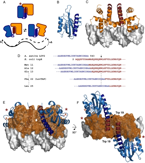
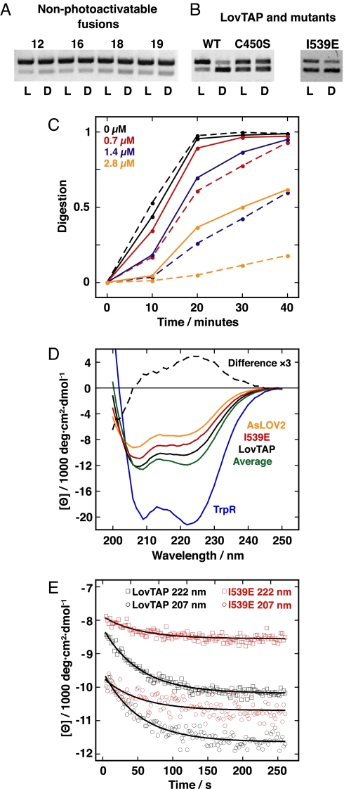
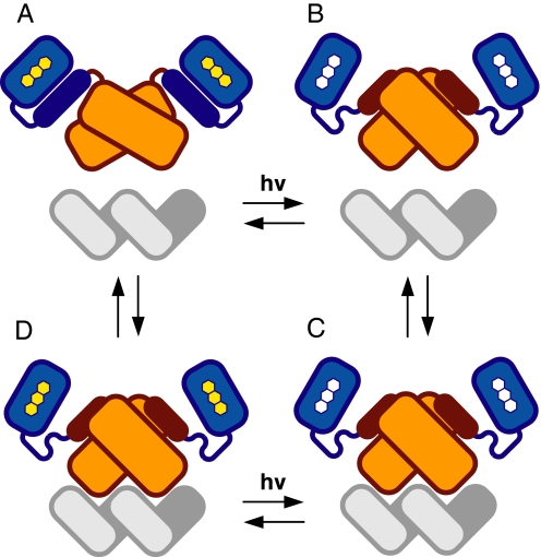
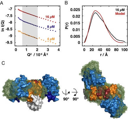
An understanding of how allostery, the conformational coupling of distant functional sites, arises in highly evolvable systems is of considerable interest in areas ranging from cell biology to protein design and signaling networks. We reasoned that the rigidity and defined geometry of an alpha-helical domain linker would make it effective as a conduit for allosteric signals. To test this idea, we rationally designed 12 fusions between the naturally photoactive LOV2 domain from Avena sativa phototropin 1 and the Escherichia coli trp repressor. When illuminated, one of the fusions selectively binds operator DNA and protects it from nuclease digestion. The ready success of our rational design strategy suggests that the helical "allosteric lever arm" is a general scheme for coupling the function of two proteins.


Conflict of interest statement

A patent application relating to this work has been filed for which D.S., K.M., and T.R.S. are coinventors.

Figures

Fig. 1.
Fig. 1.
Design of an allosteric, light-activated repressor. (A) Conceptual model of an allosteric lever arm. Joining two domains across terminal α-helices creates a bistable system in which steric overlap (red star) is relieved by the disruption of contacts between the shared helix and one or the other of the domains. A perturbation (Δ) such as ligand binding or photoexcitation alters the energy surface of the system (black line) to favor a new conformational ensemble (dashed line) with different functional properties. (B) The LOV2 domain (46) of A. sativa phototropin 1 (PDB ID code 2VOU, light blue ribbon) showing the carboxyl-terminal Jα-helix (dark blue ribbon). (C) An E. coli TrpR dimer (PDB ID code 1TRR, orange ribbon) bound to operator DNA (gray surface). The amino terminus of the protein is an α-helix (red). (D) Sequence of the Jα-helix of LOV2 through Ala 543 and of the amino terminus of TrpR beginning with Ala 2. The sequences are shown in the same colors as the models in A and B. Trp 19 of TrpR is indicated with an arrow. For this study, we created a series of constructs in which the LOV2 domain, intact through Ala 543, is fused to successive truncations of the amino-terminal helix of TrpR beginning with Met 11. (E and F) Dark-state model of LovTAP (colors same as in A and B; TrpR domain shown in orange mesh). Red stars denote regions of steric overlap. Models have been represented by using PyMOL (www.pymol.org).
Fig. 2.
Fig. 2.
Light-induced protection of operator DNA by LovTAP and associated structural changes. (A) DNA protection in the light (L) and dark (D) at 50 nM protein monomer. The examples shown are representative of all constructs except LovTAP. (B) DNA protection in the light and dark at 130 nM LovTAP monomer. (C) Light and dark activity of LovTAP. Solid lines, dark reactions; dashed lines, illuminated reactions. Digestion is the intensity ratio of the sum of product bands to the sum of the product bands plus the reactant band. The colored concentrations indicate the LovTAP monomer concentration in the reaction mixture. (D) Dark-state, far-UV CD spectra. LovTAP and the I539E mutant are shown, as are LOV2 and TrpR. The green line (Average) is the residue-weighted average of LOV2 and TrpR. The dashed line (Difference ×3) is the difference of the LovTAP spectrum and the residue-weighted average spectrum multiplied by three. (E) Kinetic recovery of CD from steady-state photoexcitation for LovTAP and the I539E mutant. Exponential fits are shown as solid lines.
Fig. 3.
Fig. 3.
Proposed mechanism for LovTAP function. The LOV domain is shown in light blue, the TrpR domain in orange, and the operator DNA in gray. The shared helix is shown in dark blue when contacting the LOV domain and in red when contacting the TrpR domain. The three-ring FMN chromophore is shown in yellow in the ground state and white when photoexcited. (A) In the dark DNA-dissociated state, the shared helix contacts the LOV domain, populating an inactive conformation of the TrpR domain. (B) Photoexcitation disrupts contacts between the shared helix and the LOV domain, populating an active conformation of the TrpR domain. (C) LovTAP binds DNA. (D) The LOV domains return to the dark state. LovTAP dissociates from the DNA, contacts between the shared helix and the LOV domain are restored, and the system returns to the initial state.
Fig. 4.
Fig. 4.
SAXS analysis of LovTAP dark-state structure. (A) Guinier plots. The shaded area indicates the range of fitting for Rg analysis (Rg·Q ≤ 1.3). (B) P(r) pair-distribution function plots. The black line is calculated from the [LovTAP] = 16 μM data. The red line is calculated from the LovTAP dark-state model, including an unstructured amino-terminal calmodulin-binding peptide (Rg = 26.9 Å). (C) Model of LovTAP compared with ab initio dark-state reconstruction from the SAXS data. The reconstruction, shown as green mesh, is manually aligned with the model of LovTAP, shown as a surface by using the same colors as in Fig. 1. DNA was not present in the experiment but is shown here in gray. The calmodulin-binding peptide is not shown.

References

    1. Bhattacharyya RP, Remenyi A, Yeh BJ, Lim WA. Domains, motifs, and scaffolds: The role of modular interactions in the evolution and wiring of cell signaling circuits. Annu Rev Biochem. 2006;75:655–680. - PubMed
    1. Pawson T, Linding R. Synthetic modular systems—Reverse engineering of signal transduction. FEBS Lett. 2005;579:1808–1814. - PubMed
    1. Pawson T, Nash P. Assembly of cell regulatory systems through protein interaction domains. Science. 2003;300:445–452. - PubMed
    1. Ponting CP, Russell RR. The natural history of protein domains. Annu Rev Biophys Biomol Struct. 2002;31:45–71. - PubMed
    1. Ostermeier M. Engineering allosteric protein switches by domain insertion. Protein Eng Des Sel. 2005;18:359–364. - PubMed

Publication types

LinkOut - more resources