. 2008 Aug 5;105(31):10895-900.
doi: 10.1073/pnas.0802702105. Epub 2008 Jul 30.

Death receptor 5 mediated-apoptosis contributes to cholestatic liver disease

Affiliations

Death receptor 5 mediated-apoptosis contributes to cholestatic liver disease

Kazuyoshi Takeda et al. Proc Natl Acad Sci U S A. .

Abstract

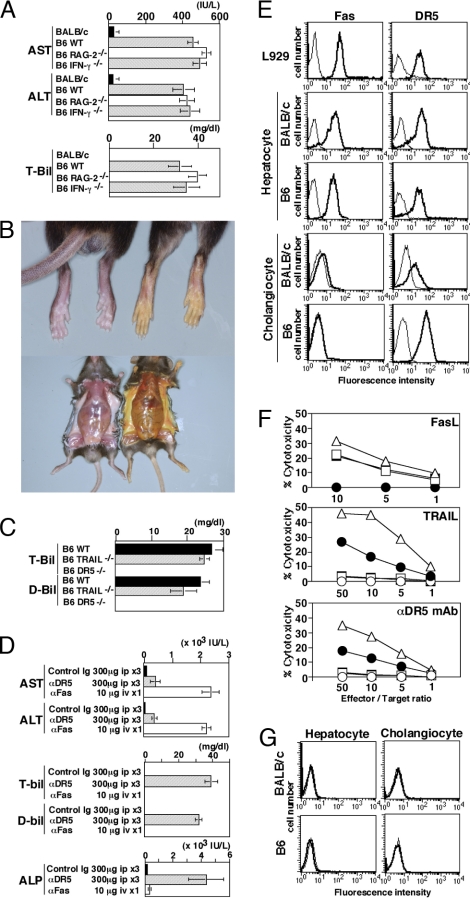
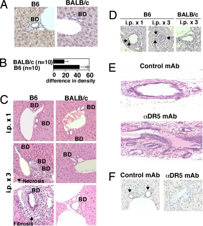
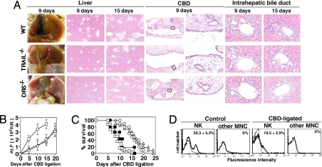
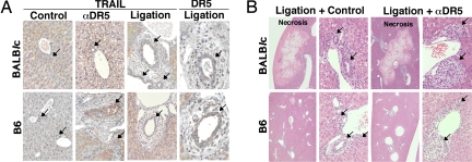
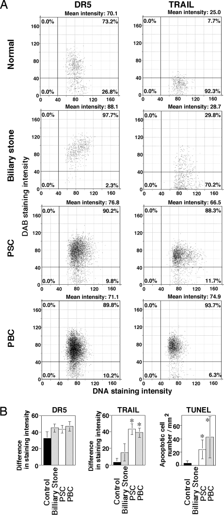
Chronic cholestasis often results in premature death from liver failure with fibrosis; however, the molecular mechanisms contributing to biliary cirrhosis are not demonstrated. In this article, we show that the death signal mediated by TNF-related apoptosis-inducing ligand (TRAIL) receptor 2/death receptor 5 (DR5) may be a key regulator of cholestatic liver injury. Agonistic anti-DR5 monoclonal antibody treatment triggered cholangiocyte apoptosis, and subsequently induced cholangitis and cholestatic liver injury in a mouse strain-specific manner. TRAIL- or DR5-deficient mice were relatively resistant to common bile duct ligation-induced cholestasis, and common bile duct ligation augmented DR5 expression on cholangiocytes, sensitizing mice to DR5-mediated cholangitis. Notably, anti-DR5 monoclonal antibody-induced cholangitis exhibited the typical histological appearance, reminiscent of human primary sclerosing cholangitis. Human cholangiocytes constitutively expressed DR5, and TRAIL expression and apoptosis were significantly elevated in cholangiocytes of human primary sclerosing cholangitis and primary biliary cirrhosis patients. Thus, TRAIL/DR5-mediated apoptosis may substantially contribute to chronic cholestatic disease, particularly primary sclerosing cholangitis.


Conflict of interest statement

The authors declare no conflict of interest.

Figures

Fig. 1.
Fig. 1.
Cholestatic liver injury in anti-DR5 mAb-treated B6 mice and sensitivity of cholangiocytes to TRAIL/DR5-induced apoptosis. (A) Serum AST, ALT, and total bilirubin (T-bil) levels of indicated mice treated with intraperitonial injections of anti-DR5 mAb (n = 10 in each group). (B) Jaundice induced by anti-DR5 mAb injections (Right), but not control Ig treatment (Left). (C) Serum T-bil and direct bilirubin (D-bil) levels after anti-DR5 mAb injections in B6 WT, DR5−/−, and TRAIL−/− mice (n = 10 in each group). (D) Serum AST, ALT, T-bil, D-bil, and ALP levels of B6 mice after repeated anti-DR5 mAb or single anti-Fas mAb injection (n = 10 in each group). (E) DR5 and Fas expression on freshly isolated B6 and BALB/c hepatocytes and cholangiocytes. Bold lines indicate the staining with anti-Fas or anti-DR5 mAb; thin lines indicate the staining with isotype-matched control Ig. (F) Sensitivity of L929 cells (triangles) and freshly isolated hepatocytes (squares) and cholangiocytes (circles) from B6 mice (filled circles) or BALB/c mice (open circles) to cytotoxicity by FasL-transfected cells, TRAIL-transfected cells, or anti-DR5 mAb-induced cytotoxicity. (G) TRAIL expression on freshly isolated B6 and BALB/c hepatocytes and cholangiocytes. Bold lines indicate the staining with anti-TRAIL mAb; thin lines indicate the staining with isotype-matched control Ig.
Fig. 2.
Fig. 2.
Cholangitis induced by anti-DR5 mAb treatment in B6 mice but not BALB/c mice. (A) Immunohistochemical examination of DR5 expression in livers of B6 and BALB/c WT mice. BD, bile duct. (B) Quantification of DR5 expression in immunohistochemistry. Sections were analyzed by using optimal densitometric mean value (MEAND), and data are represented as the mean ± SD (increased % intensity) of plural sections from indicated numbers of mice in each group. ∗, P < 0.05 compared with BALB/c mice. (C) Histological examination of livers in anti-DR5 mAb-treated B6 and BALB/c mice 4 days after one or three injections of anti-DR5 mAb. BD, bile duct. (D) Apoptosis of biliary epithelial cells demonstrated by TUNEL staining. The arrows indicate apoptotic cholangiocytes. (E) Histological examination of extrahepatic bile duct in anti-DR5 mAb- and control Ig-treated B6 mice 4 days after three injections. (F) Cholangiocytes indicated by immunohistochemical staining for cytokeratin 19. The arrows indicate intact bile ducts. Original magnification: ×20 on E and ×40 on others.
Fig. 3.
Fig. 3.
TRAIL/DR5 impairment delayed the onset of CBD ligation-induced cholestic disease. (A) Macroscopic and microscopic features of liver and bile duct in B6 WT, TRAIL−/−, and DR5−/− mice at the indicated days after CBD ligation. Original magnifications: ×5 in liver, ×2.5 and ×40 in CBD, and ×40 in intrahepatic bile duct. (B) Time kinetics of serum ALP levels in WT (squares), DR5−/− (circles) and TRAIL−/− (triangles) mice after CBD ligation. (C) Survival rate of WT (open squares), DR5−/− (open circles), TRAIL−/− (open triangles), and anti-asialo GM1 Ab-treated WT mice (filled squares) after CBD ligation. (D) TRAIL expression on liver MNC in control and CBD-ligated mice. Bold lines indicate the staining with anti-TRAIL mAb; thin lines indicate the staining with isotype-matched control Ig.
Fig. 4.
Fig. 4.
DR5 and TRAIL expression on cholangiocytes in CBD-ligated and anti-DR5 mAb-treated mice. (A) TRAIL and DR5 expression on cholangiocytes of B6 and BALB/c mice 3 days after CBD ligation, or anti-DR5 mAb-treated mice. Original magnification: ×40. (B) Cholangitis was induced with a single injection of anti-DR5 mAb in CBD-ligated mice. Original magnifications: ×5 in the images on the left and ×40 in the images on the right. The arrows indicate bile ducts.
Fig. 5.
Fig. 5.
Increased TRAIL expression and apoptosis in cholangiocytes of PSC and PBC patients. (A) Quantification of TRAIL and DR5 expression on cholangiocytes. In the representative sections (presented in Fig. S3) of each group, 324 to 6,313 of cholangiocytes were analyzed using TissueFAXS, and the results were presented as scatter grams using HistoQuest. (B) TRAIL and DR5 expression and number of apoptotic cells in cholangiocytes. Sections were analyzed using MEAND, and data are represented as the mean ± SD (increased % of intensity) of sections from plural patients in each group. ∗, P < 0.05 compared with normal and patients with CBD obstruction caused by biliary stones.

References

    1. Aggarwal BB, Shishodia S, Ashikawa K, Bharti AC. The role of TNF and its family members in inflammation and cancer: lessons from gene deletion. Current Drug Targets. 2002;1:327–341. - PubMed
    1. Locksley RM, Killeen N, Lenardo MJ. The TNF and TNF receptor superfamilies: integrating mammalian biology. Cell. 2001;104:487–501. - PubMed
    1. Ashkenazi A. Targeting death and decoy receptors of the tumour-necrosis factor superfamily. Nat Rev Cancer. 2002;2:420–430. - PubMed
    1. Janssen EM, et al. CD4+ T-cell help controls CD8+ T-cell memory via TRAIL-mediated activation-induced cell death. Nature. 2005;434:88–93. - PubMed
    1. Hamilton SE, Wolkers MC, Schoenberger SP, Jameson SC. The generation of protective memory-like CD8+ T cells during homeostatic proliferation requires CD4+ T cells. Nat Immunol. 2006;7:475–481. - PubMed

Publication types

Substances