Comparative Study
. 2008 Aug 5;105(31):10913-8.
doi: 10.1073/pnas.0801898105. Epub 2008 Jul 30.

IL-33 exacerbates antigen-induced arthritis by activating mast cells

Affiliations
Comparative Study

IL-33 exacerbates antigen-induced arthritis by activating mast cells

Damo Xu et al. Proc Natl Acad Sci U S A. .

Abstract

IL-33, a cytokine of the IL-1 family, is closely associated with type II T cell responses. Here, we report an unexpected proinflammatory role of IL-33 in inflammatory arthritis. IL-33 was expressed in synovial fibroblasts from patients with rheumatoid arthritis (RA). Expression was markedly elevated in vitro by inflammatory cytokines. Mice lacking ST2, the IL-33 receptor alpha-chain, developed attenuated collagen-induced arthritis (CIA) and reduced ex vivo collagen-specific induction of proinflammatory cytokines (IL-17, TNFalpha, and IFNgamma), and antibody production. Conversely, treatment of wild-type (WT) but not ST2(-/-) mice with IL-33 exacerbated CIA and elevated production of both proinflammatory cytokines and anti-collagen antibodies. Mast cells expressed high levels of ST2 and responded directly to IL-33 to produce a spectrum of inflammatory cytokines and chemokines in vitro. In vivo, IL-33 treatment exacerbated CIA in ST2(-/-) mice engrafted with mast cells from WT but not from ST2(-/-) mice. Disease exacerbation was accompanied by elevated expression levels of proinflammatory cytokines. Our results demonstrate that IL-33 is a critical proinflammatory cytokine for inflammatory joint disease that integrates fibroblast activation with downstream immune activation mainly via an IL-33-driven, mast-cell-dependent pathway. Thus, this IL-1 superfamily member represents a therapeutic target for RA.


Conflict of interest statement

The authors declare no conflict of interest.

Figures

Fig. 1.
Fig. 1.
Expression and regulation of IL-33 in RA synovium. (A and B) Synovial membranes from RA patients were stained with anti-IL-33 (A) and anti-ST2 (B) antibodies or isotype-matched control IgG. (C) Fibroblasts were stained for IL-33 as in A. Primary fibroblasts were purified from the synovial membrane and cultured with the cytokines indicated. (D) Fibroblasts were cultured for 16 h and IL-33 mRNA detected by RT-PCR. Pictures are representative of tissue samples from five RA patients.
Fig. 2.
Fig. 2.
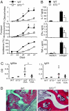
ST2−/− mice developed impaired CIA. (A) Groups of WT and ST2−/− DBA/1 mice were immunized with CII and the disease development (clinical score, footpad thickness, and incidence) was monitored. (B) DLNs of the mice in A were collected on day 27 and stimulated with CII for 3 days and the cytokine concentrations in the culture supernatant were determined by ELISA. (C) Serum anti-CII antibodies of individual samples were determined by ELISA. (D) On day 27, mice were killed and arthritic paws removed and stained with hematoxylin and eosin. Original magnification was ×50. Data are means ± SEM (*, P < 0.01 WT vs. ST2−/− mice by Mann–Whitney U test, n = 10). Data are representative of three experiments. Similar results were obtained when mice were immunized and boosted on day 21 with CII.
Fig. 3.
Fig. 3.
IL-33 exacerbated CIA. (A) Collagen-immunized and -challenged DBA/1 mice (n = 10) were injected i.p. with IL-33 (1 μg/ml) or PBS daily from days 21–25. Mice were monitored for disease progression as in Fig. 2. (B) DLN cells (2 × 10⁶ per milliliter) were stimulated with or without CII for up to 3 days and the levels of cytokines in the supernatants were determined by ELISA. (C) Serum anti-CII antibody of individual samples was determined by ELISA. (D) On day 35, mice were killed and arthritic paws removed and stained with hematoxylin/eosin or toluidine blue as in Fig. 2D. Data are mean ± SEM (*, P < 0.05; **, P < 0.01 compared with PBS controls). Data are representative of three experiments.
Fig. 4.
Fig. 4.
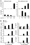
Induction of inflammatory cytokine production by IL-33 in vitro. (A) Spleen and LN cells (4 × 10⁶ per milliliter) from CIA mice were incubated in 24-well plates at 37°C for 3 h before removing nonadherent cells. Adherent and nonadherent cells (2 × 10⁶ per milliliter) were treated with IL-33 or LPS (1 μg/ml) or medium alone for 48 h. IL-6 and IL-5 concentrations in the culture supernatant were measured by ELISA. (B) BMMCs from WT or ST2−/− DBA/1 mice were cultured with IL-33 for 48 h and the cytokine and chemokine concentrations in the supernatant analyzed by Luminex as above. Data are mean ± SD and are representative of three experiments.
Fig. 5.
Fig. 5.
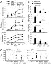
IL-33 exacerbated CIA in ST2−/− mice engrafted with mast cells from WT but not ST2−/− mice. Groups of ST2−/− mice were injected i.v. with ST2−/− or WT BMMCs (1 × 10⁷). CIA was induced 4 weeks later as in Fig. 2A. A group of WT mice was also included as positive control and immunized as engrafted ST2−/− mice. The mice were injected i.p. with IL-33 (1 μg per mouse) daily from 21 days after immunization for five days. (A) Mice were monitored for clinical score, footpad thickness, and disease incidence. (B) DLN cells were collected and cultured in vitro with collagen for 3 days, and cytokine production was determined by ELISA. (C) Serum anti-CII antibody concentration from individual mice was measured by ELISA. Data are mean ± SEM, n = 6 and are representative of two experiments.
Fig. 6.
Fig. 6.
Schematic representation of the pathogenic role of IL-33 in CIA. Tissue stroma cells including fibroblasts produce IL-33 which could be further enhanced by proinflammatory cytokines such as IL-1β and TNFα. IL-33 can promote inflammatory responses by at least three pathways. (i) IL-33 can directly activate mast cells to secrete proinflammatory cytokines which in turn cause joint inflammation. (ii) IL-1β and IL-6 secreted by IL-33-activated mast cells can promote collagen-specific Th17 cell development and function. (iii) IL-33 can also stimulate collagen-primed CD4+ T cells to produce IL-5 and IL-13 that enhance B cell activation, leading to increased IgG production. The antibodies further exacerbate joint inflammation by triggering mast cell degranulation and the formation of immune complexes with collagen.

References

    1. Schmitz JA, et al. IL-33, an interleukin-1-like cytokine that signals via the IL-1 receptor-related protein ST2 and induces T helper type 2-associated cytokines. Immunity. 2005;23:479–490. - PubMed
    1. Dinarello CA. The IL-1 family and inflammatory diseases. Clin Exp Rheumatol. 2002;20:S1–13. - PubMed
    1. Chackerian AA, et al. IL-1 receptor accessory protein and ST2 comprise the IL-33 receptor complex. J Immunol. 2007;179:2551–2555. - PubMed
    1. Ali S, et al. IL-1 receptor accessory protein is essential for IL-33-induced activation of T lymphocytes and mast cells. Proc Natl Acad Sci USA. 2007;104:18660–18665. - PMC - PubMed
    1. Xu D, et al. Selective expression and functions of interleukin 18 receptor on T helper (Th) type 1 but not Th2 cells. J Exp Med. 1998;188:1485–1492. - PMC - PubMed

Publication types