

Relationships between circadian rhythms and modulation of gene expression by glucocorticoids in skeletal muscle
- PMID: 18667713
- PMCID: PMC2576101
- DOI: 10.1152/ajpregu.90399.2008
Relationships between circadian rhythms and modulation of gene expression by glucocorticoids in skeletal muscle
Abstract
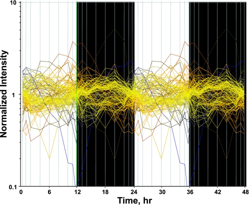
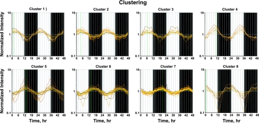
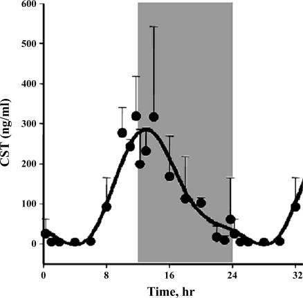
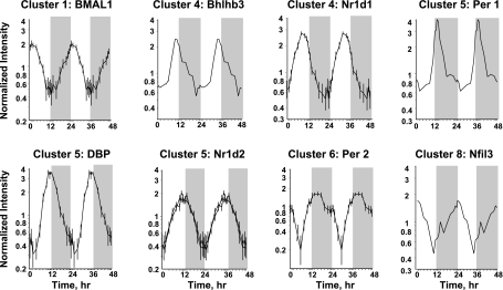
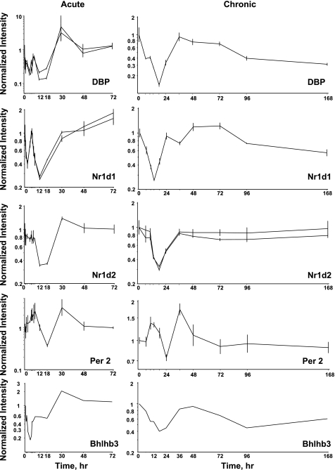
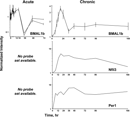
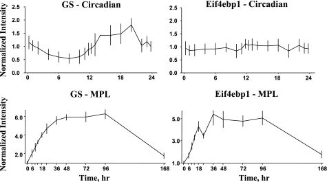
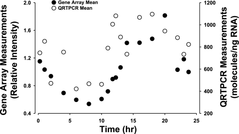
The existence and maintenance of biological rhythms linked to the 24-h light-dark cycle are essential to the health and functioning of an organism. Although much is known concerning central clock mechanisms, much less is known about control in peripheral tissues. In this study, circadian regulation of gene expression was examined in rat skeletal muscle. A rich time series involving 54 animals euthanized at 18 distinct time points within the 24-h cycle was performed, and mRNA expression in gastrocnemius muscles was examined using Affymetrix gene arrays. Data mining identified 109 genes that were expressed rhythmically, which could be grouped into eight distinct temporal clusters within the 24-h cycle. These genes were placed into 11 functional categories, which were examined within the context of temporal expression. Transcription factors involved in the regulation of central rhythms were examined, and eight were found to be rhythmically expressed in muscle. Because endogenous glucocorticoids are a major effector of circadian rhythms, genes identified here were compared with those identified in previous studies as glucocorticoid regulated. Of the 109 genes identified here as circadian rhythm regulated, only 55 were also glucocorticoid regulated. Examination of transcription factors involved in circadian control suggests that corticosterone may be the initiator of their rhythmic expression patterns in skeletal muscle.
Figures
References
-
- Adams CM, Reitz J, De Brabander JK, Fermamisco JD, Li L, Brown MS, Goldstein JL. Cholesterol and 25-hydroxycholesterol inhibit activation of SREBPs by different mechanisms, both involving SCAP and Insigs. J Biol Chem 279: 52772–52780, 2004. - PubMed
-
- Challet E, Caldelas I, Graff C, Pevet P. Synchronization of the molecular clockwork by light- and food-related cues in mammals. Biol Chem 384: 711–719, 2003. - PubMed
Publication types
MeSH terms
Substances
Grants and funding
LinkOut - more resources
Full Text Sources
Medical
Molecular Biology Databases