. 2008 Nov;79(5):878-88.
doi: 10.1095/biolreprod.108.069831. Epub 2008 Jul 30.

Restoration of the luteinizing hormone surge in middle-aged female rats by altering the balance of GABA and glutamate transmission in the medial preoptic area

Affiliations

Restoration of the luteinizing hormone surge in middle-aged female rats by altering the balance of GABA and glutamate transmission in the medial preoptic area

Genevieve S Neal-Perry et al. Biol Reprod. 2008 Nov.

Abstract

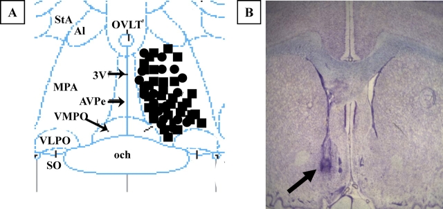
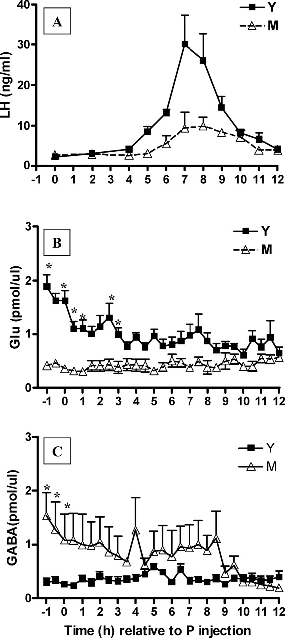
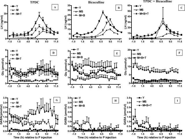
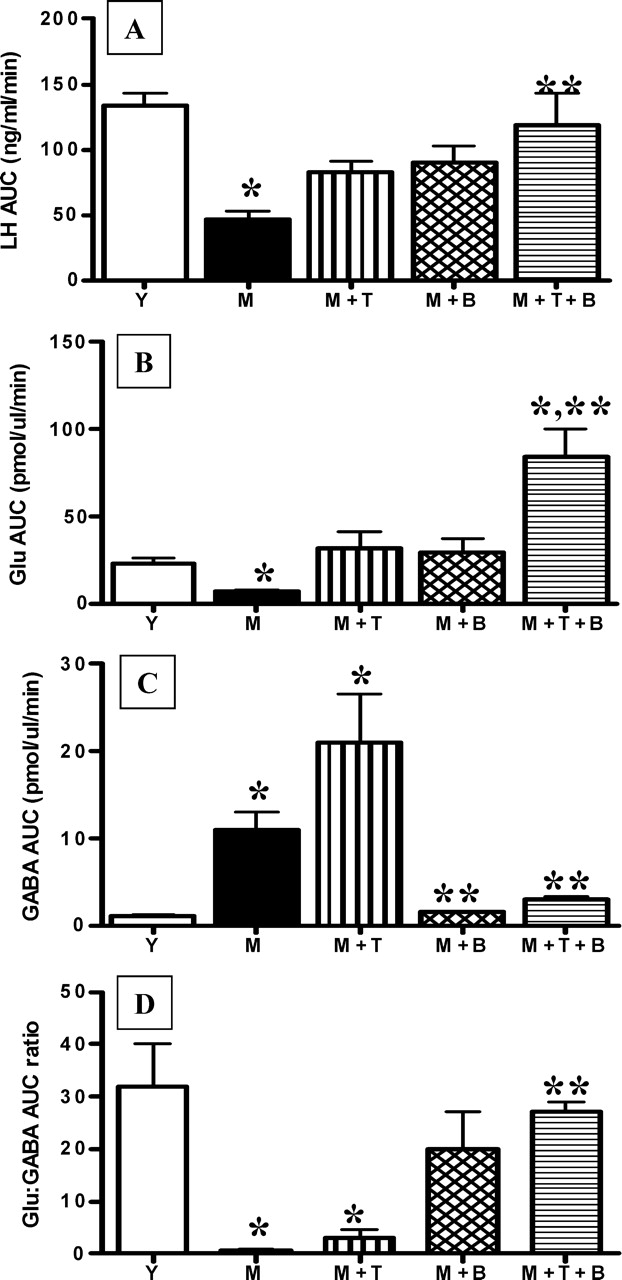
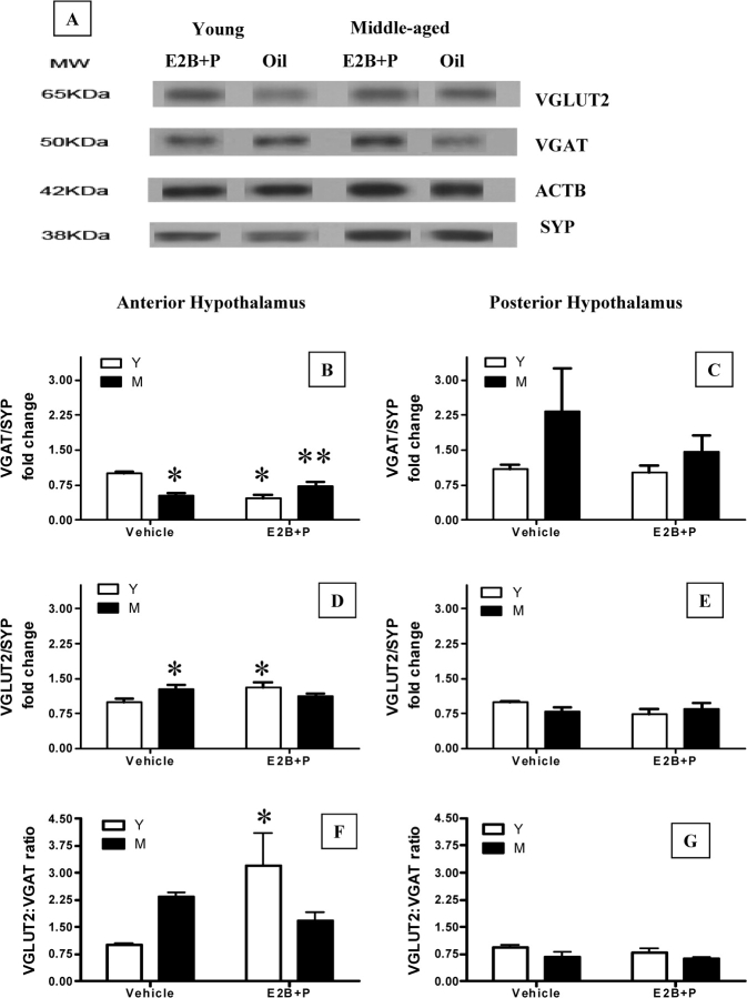
Hypothalamic glutamate and gamma-aminobutyric acid (GABA) neurotransmission are involved in the ovarian hormone-induced GnRH-LH surge in rodents. We previously reported that middle-aged rats have significantly less glutamate release in the medial preoptic area than young rats on the day of the LH surge. The present study tested the hypothesis that the delayed and attenuated LH surge in ovariohysterectomized middle-aged rats primed with ovarian steroids results from reduced hypothalamic glutamate and increased GABA(A) neurotransmission. Microdialysis results show that middle-aged rats with attenuated LH surges had reduced extracellular glutamate and increased extracellular GABA levels in the medial preoptic area compared with young rats. Blocking GABA(A) receptors with bicuculline or inhibiting synaptic glutamate reuptake with L-trans-pyrrolidine-2,4-dicarboxylic acid increased extracellular Glu in the medial preoptic area and partially restored LH surge amplitude in middle-aged rats without altering LH surge onset. Complete recovery of LH surge amplitude was observed in middle-aged rats treated with the combination of bicuculline and L-trans-pyrrolidine-2,4-dicarboxylic acid. This treatment also restored the extracellular glutamate:GABA ratio in the medial preoptic area of middle-aged rats to the level of young rats. Immunoblot analysis revealed that estradiol and progesterone treatment reduced SLC32A1(formerly known as vesicular GABA transporter) levels and increased SLC17A6 (formerly known as vesicular glutamate transporter 2) levels in the anterior hypothalamus of ovariohysterectomized young but not middle-aged rats. These data suggest that both reduced availability of glutamate and increased activation of GABA(A) receptors under estrogen-positive feedback conditions contribute to the age-related delay in onset and attenuated amplitude of the LH surge.


Figures

FIG. 1.
FIG. 1.
A) Illustration of microdialysis probes placement in the medial preoptic area. The diagram corresponds to a coronal section at approximately 0.0 mm relative to Bregma according to the atlas of Paxinos and Watson [55]. MPA, Medial preoptic area; 3V, third ventricle; och, optic chiasm; VMPO, ventromedial preoptic nucleus; VLPO, ventrolateral preoptic nucleus; AVPe, anteroventral periventricular nucleus; SO, supraoptic nucleus; Al, alar nucleus; StA, strial part preoptic nucleus; OVLT, organum vasculosum lamina terminalis. Black circles indicate young rats; black squares, middle-aged rats. B) Photomicrograph of thionin-stained coronal section showing the approximate location of a microdialysis probe. The arrow indicates the site of probe placement (original magnification ×4).
FIG. 2.
FIG. 2.
Middle-aged (M) rats exhibit a delayed and attenuated GnRH-LH surge that is accompanied by decreased extracellular Glu and increased extracellular GABA in the mPOA on the day of the GnRH-LH surge relative to young (Y) rats. Data are from GnRH-LH surges generated in Y (n = 8) and M (n = 7) rats primed with E2B and P as described in Materials and Methods. Time 0 represents the time of the P injection at 0900 h. LH (A), extracellular Glu (B), and extracellular GABA (C) levels in the mPOA. *P < 0.001. See Table 1 for additional quantitation and statistical comparisons. ul, Microliter.
FIG. 3.
FIG. 3.
Elevation of extracellular Glu and inhibition of extracellular GABA in the mPOA can increase GnRH-LH surge amplitude in middle-aged females (AI). Data are means ± SEM of young controls (Y; n = 7), middle-aged controls (M; n = 6), middle-aged controls with saline infusion at 0900 h and 1300 h (MS; n = 5), M infused at 0900 h and 1300 h with 10 μM bicuculline (M + B; n = 5), M reverse dialyzed with 10 mM TPDC (M + T; n = 6), and M treated with a combination of 10 μM bicuculline and 10 mM TPDC (M + B + T; n = 6). Time 0 is relative to the P injection at 0900 h. Each treatment is illustrated on a separate panel with the appropriate middle-aged control group (M) graphed for comparison. The same young control (Y) is depicted in each panel. See Figure 4 and Table 2 for additional quantitation and statistical comparisons.
FIG. 4.
FIG. 4.
Increasing mPOA Glu release and decreasing mPOA GABAA receptor activation with TPDC plus bicuculline rescues total LH release. Because middle-aged controls (M) and middle-aged controls with saline infusion at 0900 h and 1300 h (MS) were not significantly different, data from M are illustrated. Total LH (A), Glu (B), GABA (C), and Glu:GABA ratio (D) during the GnRH-LH surge in young control (Y; n = 8), M (n = 6), M infused at 0900 h and 1300 h with 10 μM bicuculline (M + B; n = 5), M reverse dialyzed with 10 mM TPDC (M + T; n = 6) and M treated with a combination of 10 μM bicuculline and 10 mM TPDC (M + B + T; n = 6) rats are illustrated. *Significantly different from Y (P < 0.01). **Significantly different from M (P < 0.05). AUC, Area under the curve.
FIG. 5.
FIG. 5.
Effects of steroid priming on VGAT and VGLUT2 abundance in the anterior and posterior hypothalamus. Representative Western blot gels showing abundance of synaptosomal VGAT and VGLUT2 in hypothalamic regions from vehicle and E2B + P-treated young and middle-aged rats are illustrated (A). SYP (formerly known as synaptophysin) was used as a marker for synaptosomes. ACTB (formerly known as β-actin), a cytoskeletal protein, was used as a loading control. The VGLUT2 and VGAT band densities were normalized to the SYP band density in the same sample. Normalized band density of young (Y) vehicle-treated rats (control) was assigned a value of 1 for comparison with all other groups (BG). All values are mean ± SEM of five to six independent replications (*P < 0.05 vs. young vehicle; **P < 0.05 vs. young E2B + P). M, Middle-aged control group; KDa, kilodalton.

References

    1. Burger HG, Dudley EC, Robertson DM, Dennerstein L.Hormonal changes in the menopause transition. Recent Prog Horm Res 2002; 57: 257–275.. - PubMed
    1. Park SJ, Goldsmith LT, Weiss G.Age-related changes in the regulation of luteinizing hormone secretion by estrogen in women. Exp Biol Med 2002; 227: 455–464.. - PubMed
    1. Finch CE, Felicio LS, Mobbs CV, Nelson JF.Ovarian and steroidal influences on neuroendocrine aging processes in female rodents. Endocr Rev 1984; 5: 467–497.. - PubMed
    1. Faddy MJ, Gosden RG, Gougeon A, Richardson SJ, Nelson JF.Accelerated disappearance of ovarian follicles in mid-life: implications for forecasting menopause. Hum Reprod 1992; 7: 1342–1346.. - PubMed
    1. Burger HG, Dudley EC, Hopper JL, Shelley JM, Green A, Smith A, Dennerstein L, Morse C.The endocrinology of the menopausal transition: a cross-sectional study of a population-based sample. J Clin Endocrinol Metab 1995; 80: 3537–3545.. - PubMed

Publication types