. 2008 Aug 20;27(16):2194-203.
doi: 10.1038/emboj.2008.154. Epub 2008 Jul 31.

The RNA acetyltransferase driven by ATP hydrolysis synthesizes N4-acetylcytidine of tRNA anticodon

Affiliations

The RNA acetyltransferase driven by ATP hydrolysis synthesizes N4-acetylcytidine of tRNA anticodon

Yoshiho Ikeuchi et al. EMBO J. .

Abstract

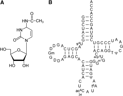
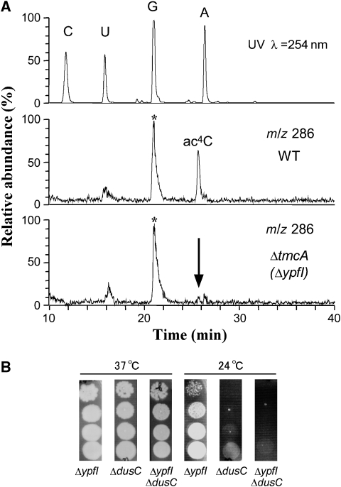
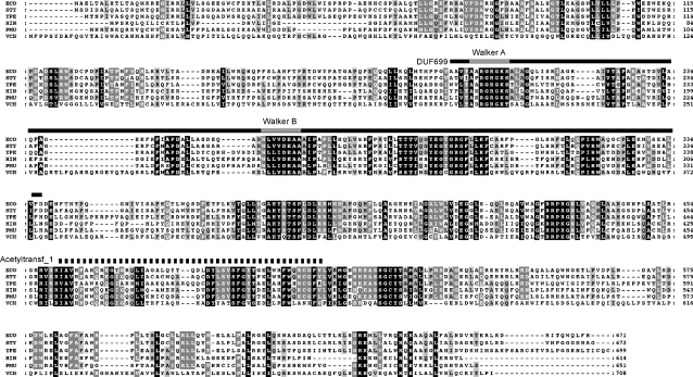
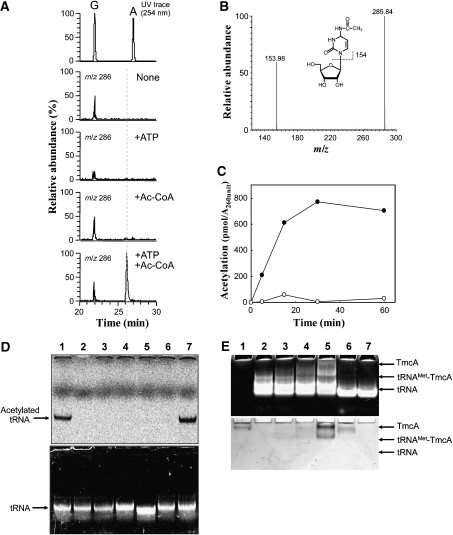
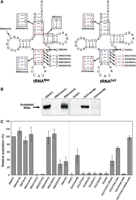
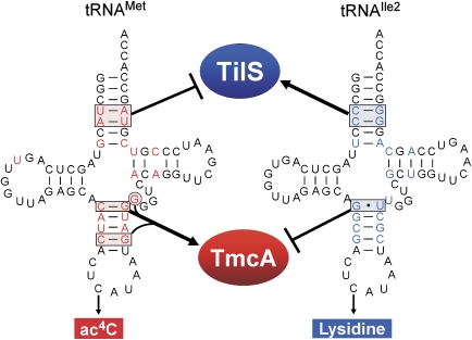
The wobble base of Escherichia coli elongator tRNA(Met) is modified to N(4)-acetylcytidine (ac(4)C), which is thought to ensure the precise recognition of the AUG codon by preventing misreading of near-cognate AUA codon. By employing genome-wide screen of uncharacterized genes in Escherichia coli ('ribonucleome analysis'), we found the ypfI gene, which we named tmcA (tRNA(Met) cytidine acetyltransferase), to be responsible for ac(4)C formation. TmcA is an enzyme that contains a Walker-type ATPase domain in its N-terminal region and an N-acetyltransferase domain in its C-terminal region. Recombinant TmcA specifically acetylated the wobble base of E. coli elongator tRNA(Met) by utilizing acetyl-coenzyme A (CoA) and ATP (or GTP). ATP/GTP hydrolysis by TmcA is stimulated in the presence of acetyl-CoA and tRNA(Met). A mutation study revealed that E. coli TmcA strictly discriminates elongator tRNA(Met) from the structurally similar tRNA(Ile) by mainly recognizing the C27-G43 pair in the anticodon stem. Our findings reveal an elaborate mechanism embedded in tRNA(Met) and tRNA(Ile) for the accurate decoding of AUA/AUG codons on the basis of the recognition of wobble bases by the respective RNA-modifying enzymes.


Figures

Figure 1
Figure 1
Chemical structure of N4-acetylcytidine (ac4C) and secondary structure of E. coli elongator tRNAMet. (A) Chemical structure of ac4C. (B) Secondary structure of E. coli elongator tRNAMet with modified nucleosides: 4-thiouridine (s4U), 2′-O-methylguanosine (Gm), dihydrouridine (D), N4-acetylcytidine (ac4C), N6-threonylcarbamoyladenosine (t6A), pseudouridine (Ψ), 7-methylguanosine (m7G), 3-(3-amino-3-carboxypropyl) uridine (acp3U) and 5-methyluridine (m5U).
Figure 2
Figure 2
Mass spectrometric analysis of total nucleosides from the ΔypfI strain and growth phenotype of the ΔypfIdusC strain. (A) LC/MS analysis of total nucleosides in the wild-type (WT) and ΔypfI strains. The top panel is the UV trace at 254 nm of the WT strain. The middle (WT) and bottom (ΔypfI) panels are mass chromatograms at m/z 286 for detecting a proton adduct (MH+) of ac4C. Absence of ac4C in ΔypfI strain is indicated by the arrow. Asterisks indicate isotopic ions of guanosine. (B) Growth property of the ΔypfIdusC strain. Each deletion strain was serially diluted (1:10 dilutions) and then spotted onto LB plates and incubated at 37 and 24 °C for 2 days.
Figure 3
Figure 3
Sequence alignment of TmcA from γ-proteobacteria. Amino-acid sequence alignment of YpfI in E. coli (ECO) and homologues in other bacteria (STY, S. typhimurium LT2; YPE, Y. pestis; HIN, H. influenzae R3021; PMU, P. multocida; and VCH, V. cholerae) were aligned. Two domains (DUF699 and Acetyltransf_1) predicted by Pfam are indicated by black solid and dotted lines, respectively. The Walker A motif is indicated by grey line. The black and grey boxes indicate a degree of sequence similarity.
Figure 4
Figure 4
Reconstitution of ac4C formation in vitro using recombinant TmcA. (A) Mass spectrometric detection of ac4C formation in the unmodified tRNAMet that was transcribed in vitro. After the reaction, the tRNAMet was digested into nucleosides and analysed by LC/MS. Mass chromatograms at m/z 286 detected ac4C, which was reconstituted in vitro in the absence of both ATP and acetyl-CoA (second panel), in the presence of ATP (third panel), in the presence of acetyl-CoA (fourth panel) or in the presence of both ATP and acetyl-CoA (bottom panel). The ac4C appeared only when both substrates were present. (B) Mass spectrum of ac4C that was reconstituted in vitro. (C) In vitro reconstitution of ac4C formation in the presence (filled circle) or absence (open circle) of ATP, which was detected by liquid scintillation counting of 14C-acetyl group. (D) In vitro reconstitution of ac4C formation under various conditions, the radioactivity of which was visualized by an imaging analyser (FLA-7000, Fujifilm). The upper and lower panels show visualized radioactivity and ethidium bromide staining. The reaction was performed in the presence of ATP (lane 1), GTP (lane 7) and ADP (lane 6), in the absence of ATP (lane 3) and TmcA (lane 2). The mutant tRNAMet, EM(C34G), whose C34 was replaced by G34 (lane 4) and tRNAIle2 (lane 5), were employed for ac4C reconstitution instead of tRNAMet. (E) Gel-retardation analysis of tRNAMet with recombinant TmcA. The polyacrylamide gel was stained by ethidium bromide (upper panel) and Coomassie brilliant blue (lower panel). Lane 1, TmcA (80 pmol); lane 2, tRNAMet; lane 3, TmcA (40 pmol) and tRNAMet; lane 4, TmcA (80 pmol) and tRNAMet; lane 5, TmcA (160 pmol) and tRNAMet; lane 6, TmcA (80 pmol) and tRNAIle2; lane 7, tRNAIle2. Amount of tRNA was 80 pmol in all conditions.
Figure 5
Figure 5
Mutation study of tRNAMet and tRNAIle2 to investigate the positive and negative determinants of ac4C formation. (A) tRNA variants based on E. coli elongator tRNAMet (left-hand side) and E. coli tRNAIle2 (right-hand side) that were used in this study. The numbering system of the tRNA is based on the tRNA compilation by Sprinzl and Vassilenko (2005). Arrows and boxes indicate the substitutions and deletions made in this study. EM and EI stand for E. coli elongator tRNAMet and E. coli tRNAIle2, respectively. (B) Detection of ac4C formation in tRNA variants visualized by the imaging analyser. (C) Relative acetylation activities of a series of tRNA variants. Radioactivity of 14C-acetylation on each tRNA was quantified by FLA-7000 system (Fujifilm). Radioactivity of ac4C in EM(WT) was standardized as 100%. A full-colour version of this figure is available at The Embo Journal Online.
Figure 6
Figure 6
Recognition and discrimination, by TmcA and TilS, of two tRNAs with CAU anticodons. Schematic depiction of tRNA recognition by TmcA in comparison with TilS. In this picture, the anticodon regions of both tRNAs are not highlighted as positive elements for both enzymes, as this region is commonly recognized by both enzymes. TmcA strongly recognizes C27-G43 in the anticodon stem of tRNAMet. In addition, C30-G40 and G44 in tRNAMet have an important function for TmcA recognition. G27·U43 in tRNAIle2 exerts an effect as a negative determinant for TmcA recognition. On the other hand, TilS strongly recognizes two consecutive base pairs, C4-G69 and C5-G68, in the acceptor stem of tRNAIle2 for lysidine formation, whereas U4-A69 and A5-U68 in tRNAMet exert an effect as negative determinants for TilS recognition.

References

    1. Alexandrov A, Chernyakov I, Gu W, Hiley SL, Hughes TR, Grayhack EJ, Phizicky EM (2006) Rapid tRNA decay can result from lack of nonessential modifications. Mol Cell 21: 87–96 - PubMed
    1. Baba T, Ara T, Hasegawa M, Takai Y, Okumura Y, Baba M, Datsenko KA, Tomita M, Wanner BL, Mori H (2006) Construction of Escherichia coli K-12 in-frame, single-gene knockout mutants: the Keio collection. Mol Syst Biol 2: 2006–2008 - PMC - PubMed
    1. Bishop AC, Xu J, Johnson RC, Schimmel P, de Crecy-Lagard V (2002) Identification of the tRNA-dihydrouridine synthase family. J Biol Chem 277: 25090–25095 - PubMed
    1. Bjork GR (1995) Biosynthesis and function of modified nucleosides. In tRNA: Structure, Biosynthesis, and Function, Söll DR, RajBhandary UL (eds), pp 165–205, Washington, DC: American Society for Microbiology
    1. Bochner BR, Ames BN (1982) Complete analysis of cellular nucleotides by two-dimensional thin layer chromatography. J Biol Chem 257: 9759–9769 - PubMed

Publication types

MeSH terms

LinkOut - more resources