. 2008 Dec;15(24):1618-22.
doi: 10.1038/gt.2008.127. Epub 2008 Jul 31.

Bioluminescent virion shells: new tools for quantitation of AAV vector dynamics in cells and live animals

Affiliations

Bioluminescent virion shells: new tools for quantitation of AAV vector dynamics in cells and live animals

A Asokan et al. Gene Ther. 2008 Dec.

Abstract

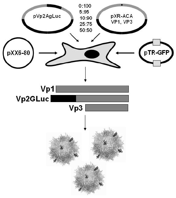
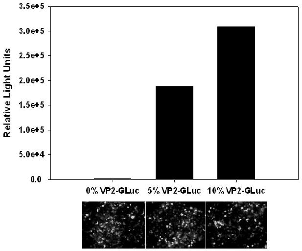
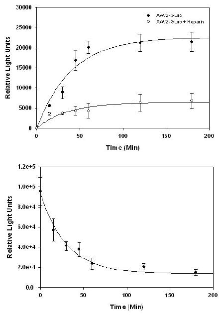
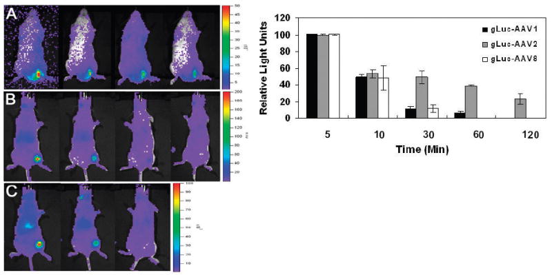
Current technologies for visualizing infectious pathways of viruses rely on fluorescent labeling of capsid proteins by chemical conjugation or genetic manipulation. For noninvasive in vivo imaging of such agents in mammalian tissue, we engineered bioluminescent Gaussia luciferase-tagged Adeno-associated viral (gLuc/AAV) vectors. The enzyme was incorporated into recombinant AAV serotypes 1, 2 and 8 capsids by fusing to the N-terminus of the VP2 capsid subunit to yield bioluminescent virion shells. The gLuc/AAV vectors were used to quantify kinetics of cell-surface-binding by AAV2 capsids in vitro. Bioluminescent virion shells displayed an exponential decrease in luminescent signal following cellular uptake in vitro. A similar trend was observed following intramuscular injection in vivo, although the rate of decline in bioluminescent signal varied markedly between AAV serotypes. gLuc/AAV1 and gLuc/AAV8 vectors displayed rapid decrease in bioluminescent signal to background levels within 30 min, whereas the signal from gLuc/AAV2 vectors persisted for over 2 h. Bioluminescent virion shells might be particularly useful in quantifying dynamics of viral vector uptake in cells and peripheral tissues in live animals.


Figures

Figure 1
Figure 1
Schematic featuring transfection protocol for production of bioluminescent AAV capsids. Plasmids pVp2gLuc and pXR-ACA are transfected at indicated ratios (total 10μg/15cm plate) along with helper Ad genes (pXX6-80) and packaging construct (pTR-GFP) in HEK 293 cells as described previously (13). Bioluminescent virion shells are generated when the Vp2gLuc protein is incorporated into AAV capsids containing Vp1 and Vp3 proteins at endogenous levels.
Figure 2
Figure 2
Functional characterization of gLuc/AAV capsids generated by transfecting 0:100; 5:95 or 10:90 ratio of plasmids pVp2gLuc and pXR2ACA. (A) Light emission from gLuc/AAV2 capsids was quantified using a Victor2® plate reader following incubation of viral fractions with coelenterazine (CLZ, 40μg/mL) substrate in 0.1M Tris buffer containing 0.6M NaCl. Average values from two different viral preparations is shown. (B) Representative fluorescence micrographs of GFP transgene expression in HEK293 cells obtained at 24hrs post-transduction with AAV2-GFP vectors (MOI 1000) containing different levels of Vp2gLuc.
Figure 3
Figure 3
Cell surface binding and uptake assays with gLuc/AAV2 capsids. (A) HelaS3 cells were incubated with gLuc/AAV2 particles (filled dots) or gLuc/AAV2 particles pre-incubated with heparin (open dots) at an MOI 10,000 at 4°C in DMEM containing 10% FBS. Aliquots of 106 cells were removed at different time intervals and cell-associated bioluminescence quantified using CLZ substrate as described earlier. (B) HelaS3 cells pre-incubated with gLuc/AAV2 particles (MOI 10,000) for 2hrs at 4°C were pelleted, washed and incubated in DMEM-FBS at 37°C. Aliquots of 107 cells were removed at different time intervals, lysed and cell-associated bioluminescence quantified using CLZ substrate as described above. All experiments were performed in triplicate. Error bars represent standard deviations.
Figure 4
Figure 4
Quantitative live animal imaging of bioluminescent gLuc/AAV capsid dynamics in Balb/C mice following intramuscular administration. Representative bioluminescent images of mice obtained using a Xenogen IVIS 100® system at 5min, 10min, 30min and 1hr post-IM administration of 5×10¹⁰ of (A) gLuc/AAV2; (B) gLuc/AAV1 and (C) gLuc/AAV8 vectors. Mice received an intravenous (tail vein) dose of 5mg/kg CLZ in MeOH/PBS immediately before IM injection gLuc/AAV particles. Light emission pseudocolor scales (total photons/sec/cm2) are shown to left of each panel. (D) Quantitation of the kinetics of light emission from left hind limb region of each mouse after IM administration of different gLuc/AAV capsids. All experiments were performed in triplicate. Error bars represent standard deviations.

References

    1. Wu Z, Asokan A, Samulski RJ. Adeno-associated virus serotypes: vector toolkit for human gene therapy. Mol Ther. 2006;14:316–327. - PubMed
    1. Warrington KH, Jr, Gorbatyuk OS, Harrison JK, Opie SR, Zolotukhin S, Muzyczka N. Adeno-associated virus type 2 VP2 capsid protein is nonessential and can tolerate large peptide insertions at its N terminus. J Virol. 2004;78:6595–6609. - PMC - PubMed
    1. Lux K, Goerlitz N, Schlemminger S, Perabo L, Goldnau D, Endell J, Leike K, Kofler DM, Finke S, Hallek M, Buning H. Green fluorescent protein-tagged adeno-associated virus particles allow the study of cytosolic and nuclear trafficking. J Virol. 2005;79:11776–11787. - PMC - PubMed
    1. Swenson ES, Price JG, Brazelton T, Krause DS. Limitations of green fluorescent protein as a cell lineage marker. Stem Cells. 2007;25:2593–2600. - PubMed
    1. Tam JM, Upadhyay R, Pittet MJ, Weissleder R, Mahmood U. Improved in vivo whole-animal detection limits of green fluorescent protein-expressing tumor lines by spectral fluorescence imaging. Mol Imaging. 2007;6:269–276. - PubMed

Publication types

MeSH terms