Comparative Study
. 2008 Aug 5;105(31):10820-5.
doi: 10.1073/pnas.0800658105. Epub 2008 Jul 31.

Huntington's disease protein contributes to RNA-mediated gene silencing through association with Argonaute and P bodies

Affiliations
Comparative Study

Huntington's disease protein contributes to RNA-mediated gene silencing through association with Argonaute and P bodies

Jeffrey N Savas et al. Proc Natl Acad Sci U S A. .

Abstract

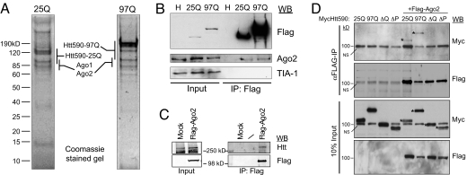
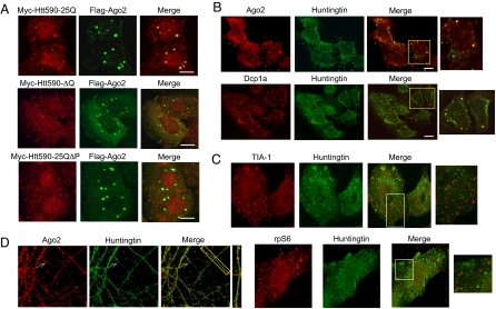
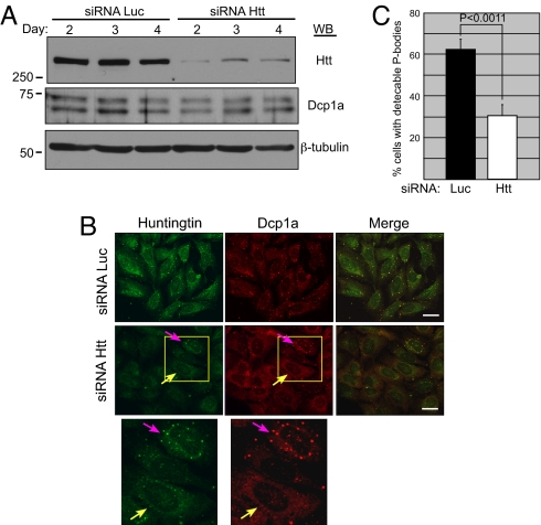
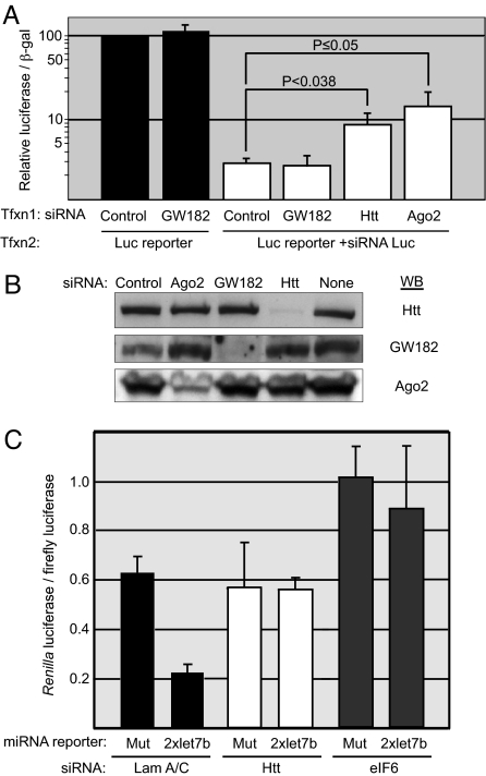
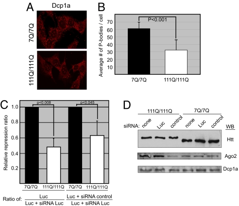
Huntington's disease is a dominant autosomal neurodegenerative disorder caused by an expansion of polyglutamines in the huntingtin (Htt) protein, whose cellular function remains controversial. To gain insight into Htt function, we purified epitope-tagged Htt and identified Argonaute as associated proteins. Colocalization studies demonstrated Htt and Ago2 to be present in P bodies, and depletion of Htt showed compromised RNA-mediated gene silencing. Mouse striatal cells expressing mutant Htt showed fewer P bodies and reduced reporter gene silencing activity compared with wild-type counterparts. These data suggest that the previously reported transcriptional deregulation in HD may be attributed in part to mutant Htt's role in post-transcriptional processes.


Conflict of interest statement

The authors declare no conflict of interest.

Figures

Fig. 1.
Fig. 1.
Isolation of Flag-tagged WT (25Q) and polyQ-expanded (97Q) Htt protein complexes from HeLa cell cytoplasmic S100 fraction. (A) Eluates from immunopurified Flag-Htt590 resolved by 10% SDS/PAGE and visualized by Coomassie blue staining. Mass spectrometric protein identifications are listed alongside the gel regions where the proteins were identified. (B) Verification of Ago2 as an Htt590 interactor. Immunoblot analysis of Flag immunoprecipitates from the indicated cell lysates probed with the indicated antibodies. (C) Cytoplasmic extract from HEK293T cells stably expressing Flag-Ago2 or Flag-vector (Mock) were immunoprecipitated with α-Flag M2 antibody and probed with α-Htt (HDB4E10, Abcam) or α-Flag antibody. (D) HeLa cells were cotransfected with Myc-Htt590–25Q, -97Q or deletion mutant (ΔQ or ΔP) and Flag-Ago2. Ago2 was immunoprecipitated with α-Flag antibody and the presence of Htt was determined by immunoblotting with α-Myc antibody. *, WT and mutant Myc-Htt590. NS: a nonspecific background band migrating at 100 kDa. Fig. S2D shows clearer separation of Myc-Htt590-ΔQ from the background band.
Fig. 2.
Fig. 2.
Htt associates with Ago2 and localizes to P bodies. (A) HeLa cells were transfected with indicated constructs as in Fig. 1D, fixed, and probed with rabbit α-Myc and mouse α-Flag primary antibodies. (Scale bar, 10 μm.) (B) U2OS cells were probed for endogenous Ago2, Dcp1a and Htt. (Scale bar, 20 μm.) Of 100 cells examined, 65% of foci showed Ago2 and Htt colocalization. Of 200 cells examined, 85% of foci showed Dcp1a and Htt colocalization. (C) U2OS cells were probed for endogenous TIA-1, rpS6, and Htt. Note that TIA-1 and rpS6 did not colocalize with endogenous Htt. (D) Primary rat hippocampal neurons were probed for endogenous Ago2 and Htt.
Fig. 3.
Fig. 3.
Htt plays a role in P body formation/maintenance. (A) U2OS cells were transfected with siRNA to Luc or Htt and the effect on Htt, Dcp1a, and β-tubulin protein was examined by immunoblotting. Quantification of Htt protein levels showed >7-fold reduction following the siRNA treatment. (B) U2OS cells transfected with Luc or Htt siRNA were examined by indirect immunofluorescence with rabbit α-Dcp1a and mouse α-Htt antibodies. (Scale bar, 30 μm.) (C) The result of three independent Luc siRNA and Htt siRNA transfections was quantified by determining the percentage of cells with detectable P-bodies after counting >200 cells per experiment.
Fig. 4.
Fig. 4.
Cells with reduced Htt levels show compromised RNA-induced gene silencing. (A) The effect of Htt knockdown on silencing of a Luc reporter with Luc siRNA. HeLa cells were first transfected (Tfxn1) with control, GW182, Htt, or Ago2 siRNA, and subsequently (Tfxn2) with SV40 promoter-Luc reporter plasmid, a plasmid encoding β-galactosidase, and Luc siRNA where indicated. The ratio of luciferase activity to β-galactosidase was measured and graphed on a log scale as the percentage of expression relative to the cells transfected with the control siRNA without Luc siRNA (set to 100%) (n = 16). (B) Immunoblot analysis of cell lysates from A. (C) The effect of Htt knockdown on the ability to silence a miRNA reporter by endogenous let-7b miRNA. HeLa cells were transfected with lamin A/C, Htt, or eIF6 siRNA, and a Renilla Luc reporter that contained human let-7b element or a mutant element. Normalized ratio of Renilla Luc to SV40-firefly Luc is presented. (n = 4).
Fig. 5.
Fig. 5.
Mouse striatal cells expressing mutant Htt have fewer P bodies and show reduced silencing activity. (A) Indirect immunofluorescence of mouse striatal cells expressing WT Htt (STHdhQ7/Q7) or mutant Htt (STHdhQ111/Q111) probed with antibodies to Dcp1a. (B) Quantification of the number of P bodies per cell performed on 100 cells. P value (paired t test) <0.001 (C) siRNA reporter assay was performed as described for Fig. 4A. The graph compares relative repression ratios in cells expressing WT (7Q/7Q) or mutant (111Q/111Q) Htt (n = 9, Left; n = 12, Right). (D) Immunoblot analysis of cell lysates prepared for the assay shown in C.
Fig. 6.
Fig. 6.
The fluorescence recovery of singly expressed RFP-Dcp1a or GFP-Ago2 within P bodies after photobleaching (mock, black) was compared with the fluorescence recovery of RFP-Dcp1a and GFP-Ago2 in HeLa cells expressing Htt590–25Q (green) or Htt590–97Q (red). The normalized recovery was fitted against a FRAP recovery model (solid lines). For clarity, not all points of the recovery data are plotted.

References

    1. The Huntington's Disease Collaborative Research Group. A novel gene containing a trinucleotide repeat that is expanded and unstable on Huntington's disease chromosomes. Cell. 1993;72:971–983. - PubMed
    1. Andrade MA, Bork P. HEAT repeats in the Huntington's disease protein. Nat Genet. 1995;11:115–116. - PubMed
    1. Li SH, Li XJ. Huntingtin and its role in neuronal degeneration. Neuroscientist. 2004;10:467–475. - PubMed
    1. Landles C, Bates GP. Huntingtin and the molecular pathogenesis of Huntington's disease. Fourth in molecular medicine review series. EMBO Rep. 2004;5:958–963. - PMC - PubMed
    1. Harjes P, Wanker EE. The hunt for huntingtin function: interaction partners tell many different stories. Trends Biochem Sci. 2003;28:425–433. - PubMed

Publication types

MeSH terms