

Review
doi: 10.1161/ATVBAHA.107.155960.
Epub 2008 Jul 31.
Assessing identity, phenotype, and fate of endothelial progenitor cells
Affiliations
- PMID: 18669889
- PMCID: PMC5244813
- DOI: 10.1161/ATVBAHA.107.155960
Item in Clipboard
Review
Assessing identity, phenotype, and fate of endothelial progenitor cells
Arterioscler Thromb Vasc Biol.
2008 Sep.
No abstract available
Figures

Common methods of “EPC” culture. Culture of colony forming unit-Hill cells (CFU-Hill, Method A, scale bar=100 um), includes a 5-day process wherein nonadherent peripheral blood mononuclear cells (PB-MNCs) give rise to the colony. Circulating angiogenic cells (CAC, Method B, scale bar=200 um) are the adherent mononuclear cells of a 4- to 7-day culture of PB-MNCs. CAC cultures typically do not display colony formation. Endothelial colony forming cells (ECFCs, Method B, scale bar=400 um) are derived from adherent PB-MNCs cultured for 6 to 21 days in endothelial conditions, and colonies display a cobblestone morphology. Only the ECFC progeny form blood vessels de novo in vivo. Images were collected using a Zeiss Axiovert 2 inverted microscope with 10×/0.25Ph1 CP-ACROMAT (CFU-EC), 32×/0.40Ph1 LD-ACROSTIGMAT (CAC), or 5× CP-ACHROMAT/0.12Ph0 (ECFC) objectives. Images were acquired using a SPOT RT color camera (Diagnostic Instruments) with the manufacturer’s software. Images cropped and scale bars added in Adobe Photoshop version 8.0. Modified from Prater DN et al. Working hypothesis to redefine endothelial progenitor cells. Leukemia. 2007;21:1142.
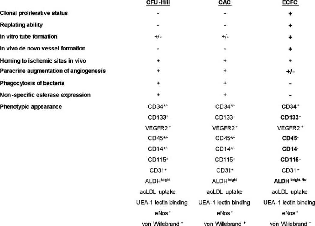
Characteristics of cells comprising the adherent population in the commonly used assays of “EPC” identification. Adherent cells that display the function are indicated by (+), that do not display the function by (−), and if the literature provides conflicting evidence (±). Those properties that distinguish cells in the ECFC assay from CFU-Hill and CAC assays are indicated in bold font. Only the ECFC and progeny display the full properties one would attribute to an EPC. VEGFR2 indicates vascular endothelial growth factor receptor 2; ALDH, aldehyde dehydrogenase; UEA-1, Ulex europeaus agglutinin-1; acLDL, acetylated low density lipoprotein; eNOS, endothelial nitric oxide synthase. The data for this figure are compiled from the articles referenced in this review.

Cord and tube formation in vitro. Although endothelial cells (A; human embryonic stem cell-derived endothelial cells, Kelly and Hirschi unpublished) form cord structures in culture, so do other unrelated cell types, such as human mammary epithelial cells (B). Only endothelial cells undergo vacuolization and form tube structures with lumens that can be demonstrated in cross-section (C; human umbilical vein endothelial cells, adapted from Davis et al).
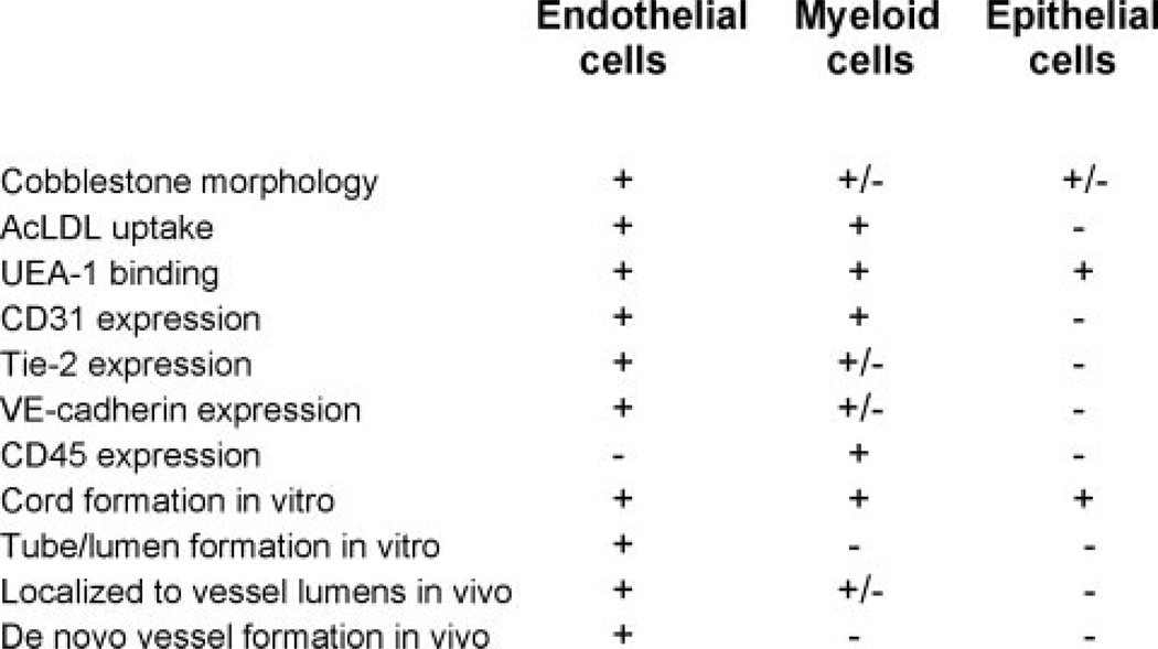
Characteristics of endothelial cells relative to myeloid and epithelial cells.
References
-
- Laubichler MD, Aird WC, Maienschein J. The endothelium in history. In: Aird WC, editor. Endothelial Biomedicine. New York: Cambridge University Press; 2007. pp. 5–19.
-
- Aird WC. Introductory essay: The endothelium in health and disease. In: Aird WC, editor. Endothelial Biomedicine. New York: Cambridge University Press; 2007. pp. 1111–1112.
-
- Bouvier CA, Gaynor E, Cintron JR, Bernhardt B, Spaet T. Circulating endothelium as an indication of vascular injury. Thromb Diath Haemorrh. 1970;40:163–168.
-
- Woywodt A, Blann AD, Kirsch T, Erdbruegger U, Banzet N, Haubitz M, Dignat-George F. Isolation and enumeration of circulating endothelial cells by immunomagnetic isolation: proposal of a definition and a consensus protocol. J Thromb Haemost. 2006;4:671–677. - PubMed
-
- Schatteman GC, Dunnwald M, Jiao C. Biology of bone marrow-derived endothelial cell precursors. Am J Physiol Heart Circ Physiol. 2007;292:H1–H18. - PubMed
Publication types
MeSH terms
Substances
Grants and funding
LinkOut - more resources
Full Text Sources
Other Literature Sources
Medical
