. 2008 Oct;39(10):2830-6.
doi: 10.1161/STROKEAHA.108.515684. Epub 2008 Jul 31.

Synergistic effect of an endothelin type A receptor antagonist, S-0139, with rtPA on the neuroprotection after embolic stroke

Affiliations

Synergistic effect of an endothelin type A receptor antagonist, S-0139, with rtPA on the neuroprotection after embolic stroke

Rui Lan Zhang et al. Stroke. 2008 Oct.

Abstract

Background and purpose: Using a model of embolic stroke, the present study tested the hypothesis that blockage of endothelin-1 with S-0139, a specific endothelin type A receptor (ET(A)) antagonist, enhances the neuroprotective effect of recombinant tissue plasminogen activator (rtPA) by suppressing molecules that mediate thrombosis and blood brain barrier (BBB) disruption induced by ischemia and rtPA.

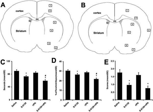
Methods: Rats (n=104) subjected to embolic middle cerebral artery (MCA) occlusion were randomly divided into 1 of 4 infusion groups with 26 rats per group: (1) the control group in which rats were administered saline, (2) the monotherapy rtPA group in which rtPA was intravenously administered at a dose of 10 mg/kg 4 hours after MCA occlusion, (3) the monotherapy S-0139 group in which S-0139 was intravenously given 2 hours after MCA occlusion, and (4) the combination of rtPA +S-0139 group in which S-0139 and rtPA were given 2 and 4 hours after MCA occlusion, respectively. Measurements of infarct volume and parenchymal hemorrhage, behavioral outcome, and immunostaining were performed on rats euthanized 1 and 7 days after stroke.

Results: The combination therapy of S-0139 and rtPA significantly (P<0.01) reduced infarct volume (24.8+/-0.9% versus 33.8+/-1.5% in control) and hemorrhagic area (7.1+/-6.1 microm(2) versus 36.5+/-19.2 microm(2) in control) and improved functional recovery compared with control saline-treated animals. Immunostaining analysis revealed that the combination therapy had the synergistically suppressed ischemia- and rtPA-induced ICAM-1, protease-activated receptor 1 (PAR-1), as well as accumulation of platelets in cerebral microvessels. Furthermore, the combination treatment synergistically reduced loss of laminin, ZO1, and occludin in cerebral vessels.

Conclusions: These data suggest that S-0139 provides the neuroprotection by suppressing ischemia- and rtPA-triggered molecules that evoke thrombosis and BBB disruption.


Figures

Figure 1
Figure 1
A schematic of coronal sections shows 6 (A) and 7 (B) areas where images were acquired for analysis of thrombocytes and vascular integrity, respectively. The effects of S-0139 on behavioral tests (C to E). Monotherapy of S-0139 given 2 hours after stroke and combination therapy of S-0139 given 2 hours and rtPA administered 4 hours after stroke improve neurological function measured by adhesive removal test (C), foot-fault test (D), and mNSS (E) 7 days after MCA occlusion. *P<0.05 vs the saline group and #P<0.05 vs monotherapy of S-0139 group. N=20 rats/group. CC indicates corpus callosum; lv, lateral ventricle.
Figure 2
Figure 2
Immunostaining shows ICAM-1 immunoreactive vessels in the ischemic hemisphere from representative rats treated with rtPA alone (A through C) and S-0139 in combination with rtPA (D through F). C and F indicate the cortex. B and E represent the striatum. G, Quantitative data of ICAM-1 immunoreactive vessels (n=6 per group, # vs saline and * vs rtPA). Bar=25 µm (B to F). Monotherapy of rtPA was administered 4 hours after stroke, whereas combination therapy of S-0139 and rtPA was given 2 and 4 hours after stroke, respectively.
Figure 3
Figure 3
Photomicrographs show PAR-1 (A and B) and platelet (C and D) immunoreactive vessels in the ischemic boundary of the cortex (area 2 of Figure 1A) from representative rats treated with rtPA alone (A and C) or S-0139 in combination with rtPA (B and D). E and F, Quantitative data of PAR-1 and thrombocyte immunoreactive vessels (n=6 per group), respectively. Please see methods for details of quantification of PAR-1 and thrombocyte immunoreactive vessels. Bar=25 µm (A to D). Monotherapy of rtPA was administered 4 hours after stroke, whereas combination therapy of S-0139 and rtPA was given 2 and 4 hours after stroke, respectively.
Figure 4
Figure 4
Photomicrographs show collagen IV (A to C) and laminin (E to G) immunoreactive vessels in the cortex of homologous areas of the contralateral hemisphere (A and E) and in the ischemic boundary of the cortex (areas 1 and 2 of Figure 1B) from representative rats treated with rtPA alone (B and F) or S-0139 in combination with rtPA (C and G). D and H, Quantitative data of percentage of collagen IV and laminin, respectively, positive vessels in the ipsilateral hemisphere vs the contralateral hemisphere (n=6 per group). Bar=20 µm (A to G). Monotherapy of rtPA was administered 4 hours after stroke, whereas combination therapy of S-0139 and rtPA was given 2 and 4 hours after stroke, respectively.
Figure 5
Figure 5
Photomicrographs show ZO1 (A to D) and occludin (F to I) immunoreactive vessels in the cortex of homologous areas of the contralateral hemisphere (A and F) and in the ischemic boundary of the cortex (areas 1 and 2 of Figure 1B) from representative rats treated with saline (B and G), rtPA alone (C and H), or S-0139 in combination with rtPA (D and I). E and J, Quantitative data of percentage of ZO1 and occludin, respectively, positive vessels in the ipsilateral hemisphere vs the contralateral hemisphere. (n=6 per group). Bar=20 µm (A to I). Monotherapy of rtPA was administered 4 hours after stroke, whereas combination therapy of S-0139 and rtPA was given 2 and 4 hours after stroke, respectively.

References

    1. Inoue A, Yanagisawa M, Kimura S, Kasuya Y, Miyauchi T, Goto K, Masaki T. The human endothelin family: Three structurally and pharmacologically distinct isopeptides predicted by three separate genes. Proc Natl Acad Sci U S A. 1989;86:2863–2867. - PMC - PubMed
    1. Stanimirovic DB, Yamamoto T, Uematsu S, Spatz M. Endothelin-1 receptor binding and cellular signal transduction in cultured human brain endothelial cells. J Neurochem. 1994;62:592–601. - PubMed
    1. Narushima I, Kita T, Kubo K, Yonetani Y, Momochi C, Yoshikawa I, Ohno N, Nakashima T. Highly enhanced permeability of blood-brain barrier induced by repeated administration of endothelin-1 in dogs and rats. Pharmacol Toxicol. 2003;92:21–26. - PubMed
    1. Matsuo Y, Mihara S, Ninomiya M, Fujimoto M. Protective effect of endothelin type a receptor antagonist on brain edema and injury after transient middle cerebral artery occlusion in rats. Stroke. 2001;32:2143–2148. - PubMed
    1. Volpe M, Cosentino F. Abnormalities of endothelial function in the pathogenesis of stroke: The importance of endothelin. J Cardiovasc Pharmacol. 2000;35:S45–S48. - PubMed

Publication types

MeSH terms